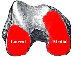
Screw Home Mechanism of The Knee Joint
Definition[edit | edit source]
Screw home mechanism (SHM) of knee joint is a critical mechanism that play an important role in terminal extension (last 15 degress) of the knee [1][2][3][4][5][6]. Knee joint is a hingle type, uniaxial joint that alows flexion and extension movements. Hovewer there is a slight rotation in last 15 degrees of knee extention due to inequality of articlar surface of femur condlyles.
SHM is a result of Vastusm Medialis Obliquus and Vastus Lateralis Obliquus mucle function. Functionally it increases the mechanical advantage of kneee joint to perform full terminal extention then locks the knee joint[7]. Popliteus muscle unloks the knee.
In most musculoskeletal problems of knee loss of terminal extention is a common problem and SHM have a clinical importance[8][9][6].
Physiological/Mechanical Movement Patern[edit | edit source]
Mechanical Paradox: Articular surface of medial condyle of femur is greater than the articular surface of later condyle.
Physiological Movement: Both medial and lateral condyle moves on articular surface of tibia (medial/lateral meniscus)
Kinetic Chain: Open kinetic chain (foot and calf freely moves)
Movement: Terminal extantion of the knee
Screw Home Movement: During extantion when all articular surface of lateral condyle is used by roll movement there are still unused articular surface on medial condyle. Femur glides posteriorly on tibia to use full articular surface of medial condyle. Then knee is locked by vastus medialis obliquus.
Clinical Relevelance[edit | edit source]
In most of the knee problems genrally there is an insufficeny on terminal extention and vastus medialis obliquus function. Restoration of terminal extantion is an important goal of rehabilitation program. Terminal extention exercies like interventions have important contribution on restoration of screw home movement /terminal extention.
References[edit | edit source]
- ↑ Goodfellow J, O'Connor J. The mechanics of the knee and prosthesis design. J Bone Joint Surg Br. 1978;60(3):358–369.
- ↑ Bytyqi D, Shabani B, Lustig S, Cheze L, Karahoda Gjurgjeala N, Neyret P. Gait knee kinematic alterations in medial osteoarthritis: three dimensional assessment. Int Orthop. 2014;38(6):1191–1198.
- ↑ Ishii Y, Terajima K, Terashima S, Koga Y. Three-dimensional kinematics of the human knee with intracortical pin fixation. Clin Orthop Relat Res. 1997;(343):144–150.
- ↑ https://www.ncbi.nlm.nih.gov/pubmed/12135550
- ↑ Asano T, Akagi M, Tanaka K, Tamura J, Nakamura T. In vivo three-dimensional knee kinematics using a biplanar imagematching technique. Clin Orthop Relat Res. 2001;(388):157–166
- ↑ 6.0 6.1 Kim, H. Y., Kim, K. J., Yang, D. S., Jeung, S. W., Choi, H. G., & Choy, W. S. (2015). Screw-Home Movement of the Tibiofemoral Joint during Normal Gait: Three-Dimensional Analysis. Clinics in orthopedic surgery, 7(3), 303–309. doi:10.4055/cios.2015.7.3.303
- ↑ Bevilaqua-Grossi D1, Monteiro-Pedro V, de Vasconcelos RA, Arakaki JC, Bérzin F. The effect of hip abduction on the EMG activity of vastus medialis obliquus, vastus lateralis longus and vastus lateralis obliquus in healthy subjects. J Neuroeng Rehabil. 2006 3;3:13.
- ↑ Udagawa K, Niki Y, Enomoto H, Toyama Y, Suda Y. Factors influencing graft impingement on the wall of the intercondylar notch after anatomic double-bundle anterior cruciate ligament reconstruction. Am J Sports Med. 2014;42(9):2219–2225.
- ↑ Lee TQ. Biomechanics of hyperflexion and kneeling before and after total knee arthroplasty. Clin Orthop Surg. 2014;6(2):117–126.


