Scapular balance angle
Invalid title.
Purpose[edit | edit source]
The condition of altered scapular mechanics and motion is called ‘scapular dyskinesis’, where ‘dys’ indicates alteration and ‘kinesis’ indicates motion. it can be found in healthy individuals or be responsible for a syndrome characterised by several symptoms called SICK. (Scapular malposition, Inferior medial border prominence, Coracoid pain and malposition, and DysKinesis of scapular motion)[1].
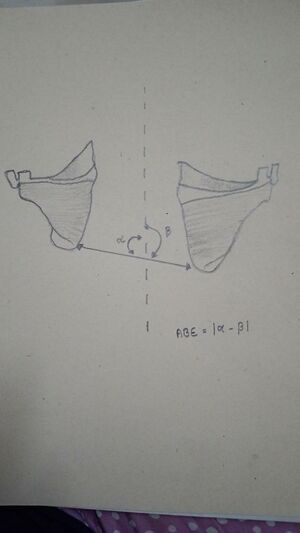
scapular balance angle (SBA) is defined as “the difference between the angles formed by the line joining the 2 inferior angles of the scapula with the vertical line passing through the spine”[2]
The most common pathologies that are associated with some form of scapular Dyskinesis are: (1) Acromio clavicular instability, (2) shoulder impingement, (3) rotator cuff injuries, (4) Glenoid labrum injuries, (5) clavicle fracture and (6) nerve-related.
Technique[edit | edit source]
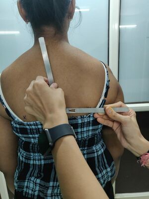
- The Scapular balance angle measurement is obtained with the patient standing on both bare feet, with arms, pelvis, and heels together.
- Next, the inferior angle of the scapula was marked bilaterally and the line connecting these marks and another vertical line between the C7 and T9---T10 spinous processes were drawn.
- Lastly, the angles formed by the line joining both inferior angles of the scapula with the vertical line running through the spine were measured with the goniometer.
- The SBA was defined as ‘‘the difference between the angles formed by the line joining the 2 inferior angles of the scapula with the vertical line passing through the spine. SBA greater than 7 ◦ would entail the diagnosis of scapular dyskinesia[2].
- Manual measurement of the SBA is reproducible at an intra observer (ICC: 0.87) and inter observer (ICC: 0.84) level.[2]
- This criterion presented a sensitivity of 72.73%, specificity of 90.91% and odds ratio of 8[2].


Evidences[edit | edit source]
- Manual measurement of the SBA is reproducible at an intra-observer (ICC: 0.87) and inter-observer (ICC: 0.84) level.
- This criterion presented a sensitivity of 72.73%, specificity of 90.91% and odds ratio of 8.
Physiotherapy Management[3][4][5][6][7][edit | edit source]
Conservative treatment in SD cases aims to restore scapular retraction, posterior tilt, and external rotation. Specific exercises for scapular rehabilitation include flexibility exercises to decrease scapular traction, and scapular stabilization exercises to optimize scapular kinematics.
Scapular stabilization exercises, based on stretching and strengthening, aim to improve muscle strength and joint position sense The serratus anterior and trapezius muscles act as scapular stabilizers.
The serratus anterior plays an essential role in determining scapular external rotation and posterior tilt, and the lower trapezius helps to stabilize the scapular position.
Scapular stabilization exercises are based on closed and open kinetic chain exercises, including push-ups on a stable or unstable surface, lawnmower exercises, and resisted scapular retraction.
References[edit | edit source]
- ↑ López-Vidriero E, López-Vidriero R, Rosa LF, et al. Scapular dyskinesis: related pathology. International Journal of Orthopaedics 2015;2(1):191-5.
- ↑ 2.0 2.1 2.2 2.3 2.4 2.5 Contreras J, Gil D, de Dios Errázuriz J, Ruiz P, Díaz C, Águila P, et al. Valores de referencia del ángulo de balance escapular en población sana. Rev Esp Cir Ortop Traumatol. 2014;58:24---30.
- ↑ Giuseppe LU, Laura RA, Berton A, Candela V, Massaroni C, Carnevale A, Stelitano G, Schena E, Nazarian A, DeAngelis J, Denaro V. Scapular dyskinesis: from basic science to ultimate treatment. International journal of environmental research and public health. 2020 Apr;17(8):2974.
- ↑ Umehara J, Nakamura M, Nishishita S, Tanaka H, Kusano K, Ichihashi N. Scapular kinematic alterations during arm elevation with decrease in pectoralis minor stiffness after stretching in healthy individuals. Journal of shoulder and elbow surgery. 2018 Jul 1;27(7):1214-20.
- ↑ Başkurt Z, Başkurt F, Gelecek N, Özkan MH. The effectiveness of scapular stabilization exercise in the patients with subacromial impingement syndrome. Journal of back and musculoskeletal rehabilitation. 2011 Jan 1;24(3):173-9.
- ↑ Turgut E, Duzgun I, Baltaci G. Effects of scapular stabilization exercise training on scapular kinematics, disability, and pain in subacromial impingement: a randomized controlled trial. Archives of physical medicine and rehabilitation. 2017 Oct 1;98(10):1915-23.
- ↑ Morais N, Cruz J. The pectoralis minor muscle and shoulder movement-related impairments and pain: Rationale, assessment and management. Physical Therapy in Sport. 2016 Jan 1;17:1-3.


