Pronator Teres: Difference between revisions
No edit summary |
No edit summary |
||
| Line 42: | Line 42: | ||
=== Trigger points === | === Trigger points === | ||
{{#ev:youtube|coyzeDwD9_k}} | {{#ev:youtube|coyzeDwD9_k}}<ref>TrPTherapist. The Pronator Teres Trigger Point. Available from: https://www.youtube.com/watch?v=coyzeDwD9_k [last accessed 11/11/2021] </ref> | ||
== Assessment == | == Assessment == | ||
| Line 66: | Line 66: | ||
=== Manual therapy === | === Manual therapy === | ||
{{#ev:youtube|3tKlRyC7KUU}} | {{#ev:youtube|3tKlRyC7KUU}}<ref>Whitney Lowe. Active Engagement on Pronator Teres. Available from: https://www.youtube.com/watch?v=3tKlRyC7KUU [ last accessed 11/11/2021] </ref> | ||
== See also == | == See also == | ||
| Line 75: | Line 75: | ||
*[[Cubital Fossa]] | *[[Cubital Fossa]] | ||
==Resources== | ==Resources== | ||
{{#ev:youtube|sQAUX_5SjRc}} | {{#ev:youtube|sQAUX_5SjRc}}<ref>nabil ebraheim. Anatomy Of The Pronator Teres Muscle - Everything You Need To Know - Dr. Nabil Ebraheim. Available from: https://www.youtube.com/watch?v=sQAUX_5SjRc [last accessed 11/11/2021] </ref> | ||
= References = | = References = | ||
Revision as of 00:53, 12 November 2021
This article is currently under review and may not be up to date. Please come back soon to see the finished work! (12/11/2021)
Top Contributors - Peter Zatezalo, Ilona Malkauskaite, Joao Costa, Kirenga Bamurange Liliane, Evan Thomas and Ewa JaraczewskaPeter Zatezalo, DPT
Description[edit | edit source]
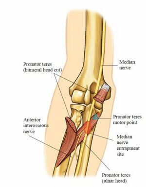
The pronator teres muscle is a long, round muscle that is located on the anterior aspect of the forearm. This muscle has two different points of origin: the humeral head and the ulnar head. [1] The median nerve commonly passes between the two heads of the pronator teres (83% of arms) where it is at risk for compression.[2] The pronator teres forms the medial border of the Cubital Fossa, as illustrated in the image.
Origin[edit | edit source]
Origin of Humeral Head: Immediately above the medial epicondyle of the humerus, common flexor tendon and deep antebrachial fascia.
Origin of Ulnar Head: Medial side of the coronoid process of the ulna[3].
Insertion[edit | edit source]
Middle of the lateral surface of the radius.[3]
Nerve[edit | edit source]
Median nerve, C6 and C7.[3]
Artery[edit | edit source]
Ulnar artery, anterior recurrent ulnar artery.[4]
Function[edit | edit source]
Pronator teres pronates the forearm and assists in flexion of the elbow joint.[3]It acts synergistically with the pronator quadratus. If the elbow is fully flexed, then the muscle fibers are shortened and less able to produce force.
Synergist: Pronator Quadratus
Antagonist: Supinator, Biceps Brachii
Clinical relevance[edit | edit source]
Nerve Entrapment[edit | edit source]
The median nerve typically runs between the two heads of the pronator teres, making it a possible site of nerve entrapment. There will likely be pain and paresthesia in the distribution of the median nerve.
This entrapment can present similarly to carpal tunnel because the median nerve is affected. If pronator syndrome is present, the individual should have a negative Phalen’s Test, negative Tinel’s Test at the wrist (and a positive Tinel sign at the pronator teres), and likely pain with resisted pronation. The Pronator Teres Syndrome Test would likely be positive. In addition, there may be history of repetitive pronation/supination such as with a person who plays racquet sports.
Trigger points[edit | edit source]
Assessment[edit | edit source]
Palpation[edit | edit source]
The pronator arises from the common flexor tendon at the medial epicondyle of the humerus. It passes obliquely from medial to lateral and forms the medial border of the cubital fossa. To palpate it, feel the antecubital fossa, most slightly medially, and pronate/supinate the forearm.
Strength[edit | edit source]
Patient is supine or sitting. The elbow should be held against the patients side or be stabilized by the examiner to avoid any shoulder abduction movement.
Test: Pronation of the forearm with the elbow partially flexed. Pressure: At the lower forearm , above the wrist (to avoid twisting the wrist), in the direction of supinating the forearm. [3]
Treatment[edit | edit source]
Strengthening[edit | edit source]
| [7] | [8] |
Manual therapy[edit | edit source]
See also[edit | edit source]
Resources[edit | edit source]
References[edit | edit source]
- ↑ https://study.com/academy/lesson/pronator-teres-definition-function-nerve-supply.html (accessed 20 July 2018).
- ↑ Doyle, J. Surgical Anatomy of the Hand and Upper Extremity. Philadelphia: Lippincott, Williams, and Wilkins. 2003
- ↑ 3.0 3.1 3.2 3.3 3.4 Kendall F, McCreary E, Provance P,Rodgers M,Romani W. Muscles:Testing and function with posture and pain. 5th ed. Philadelphia: Lippincott Williams & Wilkins, 2005.
- ↑ https://rad.washington.edu/muscle-atlas/pronator-teres/ (accessed 20 July 2018).
- ↑ TrPTherapist. The Pronator Teres Trigger Point. Available from: https://www.youtube.com/watch?v=coyzeDwD9_k [last accessed 11/11/2021]
- ↑ Black River and Bootsma Education. Muscle Palpation - Pronator Teres. Available from: https://www.youtube.com/watch?v=yqkvnxqV18Q [last accessed 11/11/2021]
- ↑ The Pronator. The Pronator. The Pronator: Forearm Pronation and Supination. Available from: https://www.youtube.com/watch?v=qG9jjLqYvdA [last accessed 11/11/2021]
- ↑ Bob and Brad. Little Known Exercises That Drastically Increase Grip Strength. Available from: https://www.youtube.com/watch?v=qGaFpQP1n9o [last accessed 11/11/2021]
- ↑ Whitney Lowe. Active Engagement on Pronator Teres. Available from: https://www.youtube.com/watch?v=3tKlRyC7KUU [ last accessed 11/11/2021]
- ↑ nabil ebraheim. Anatomy Of The Pronator Teres Muscle - Everything You Need To Know - Dr. Nabil Ebraheim. Available from: https://www.youtube.com/watch?v=sQAUX_5SjRc [last accessed 11/11/2021]


