Navicular stress syndrome: Difference between revisions
Sazia Queyam (talk | contribs) mNo edit summary |
Sazia Queyam (talk | contribs) mNo edit summary |
||
| Line 4: | Line 4: | ||
=== INTRODUCTION === | === INTRODUCTION === | ||
The Navicular is an intermediate tarsal bone on the medial side of the foot<ref>D.Richard, V.Wayne, M. Adam, Gray’s Anatomy for Students. Spain: Elsevier Publishers, 2005</ref>. | The Navicular is an intermediate tarsal bone on the medial side of the foot<ref>D.Richard, V.Wayne, M. Adam, Gray’s Anatomy for Students. Spain: Elsevier Publishers, 2005</ref>. | ||
[[File:Foot accessory navicular CLINICAL ANATOMY 1 anat01.jpg|left|Navicular Bone]][[File:Navicular Bone Articulation.gif|none|The left navicular. Antero-lateral view.]] [[File:Navicular bone articulation PM.gif| | [[File:Foot accessory navicular CLINICAL ANATOMY 1 anat01.jpg|left|Navicular Bone]][[File:Navicular Bone Articulation.gif|none|The left navicular. Antero-lateral view.]] [[File:Navicular bone articulation PM.gif|left|The left navicular. Postero-medial view.]] | ||
Revision as of 19:55, 15 December 2018
-----PAGE IS UNDER CONSTRUCTION----
INTRODUCTION[edit | edit source]
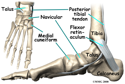
The Navicular is an intermediate tarsal bone on the medial side of the foot[1].
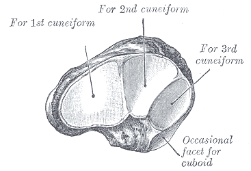
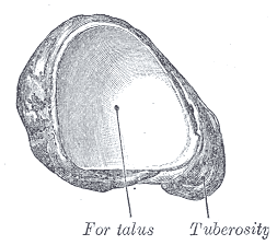
Its name (os naviculare pedis; scaphoid bone) derives from the human bone's resemblance to a small boat. It articulates with four bones: the talus and the three cuneiforms; occasionally with a fifth, the cuboid.
CLINICALLY RELEVANT ANATOMY[edit | edit source]
REFERENCES[edit | edit source]
- ↑ D.Richard, V.Wayne, M. Adam, Gray’s Anatomy for Students. Spain: Elsevier Publishers, 2005


