Morel-Lavallée lesion
Definition[edit | edit source]
A Morel-Lavallée lesion (MLL) was first described in 1853[1] [2]. It is a closed soft-tissue degloving injury[1][3][4][5][6] that usually occurs after blunt trauma[1][2][3][5]. In recent literature, it can also be referred to as Morel-Lavallée seroma or effusion, post-traumatic soft tissue cysts or post-traumatic extravasations[2].
Epidemiology and aetiology[edit | edit source]
These injuries are uncommon[2] and there is no consensus on the ratio of men to women. One source reported a 2:1 ratio[2] while another reported a 1:1 ratio[7].
These injuries occur due to blunt trauma after:
MLL can also be iatrogenic e.g. after abdominal liposuction or mammoplasty[1][2][6]
Pathophysiology[edit | edit source]

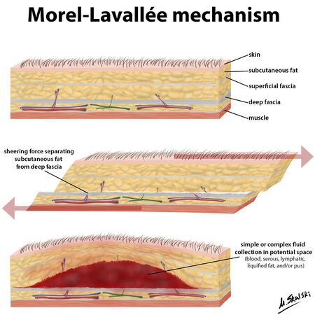
MLL occurs due to shearing forces which separate the skin and subcutaneous tissue from the deep fascia, causing a potential space[1][2][4][5][8][10][11]. Damage to the lymphatic and blood vessels leads to an accumulation of blood and lymph[1][3][4][8][10] and necrotic fat[1][3][8][12] in the potential space, causing a haematoma or seroma[10]. Blood will start to be reabsorbed over time leaving a serosanguinous fluid surrounded by a haemosiderin layer[2]. Inflammation is then induced by the haemosiderin layer leading to a fibrous capsule[2][11]. This fibrous capsule prevents more fluid reabsorption, initiating a chronic MLL[3].
MLL are often associated with pelvic or acetabular fractures but can also occur without a fracture[9].
Secondary risk factors for an MLL include female gender and BMI of over 25[1].
Clinical Presentation[edit | edit source]

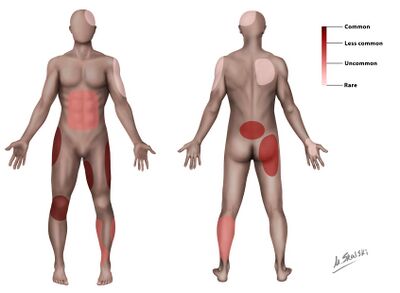
MLL occurs most commonly over the greater trochanter (>60% of cases)[1][3] [5][6][8][11], proximal femur[1][2][5], buttock[2][3][5], knee[3][5][6][11] and in rare cases, the lumbar region[2][3][6][11]. It can also occur at the scapula[2][6]. Delayed presentation (months or years) can occur in up to ⅓ of patients[1][8]. The most common signs and symptoms include:
- Compressible, fluctuant swollen area[1][2][5][8][9]. The fluctuant swelling is an essential clinical characteristic[9].
- Pain[1][2][4][8]
- Stiffness[5][9]
- Cutaneous anaesthesia or hypothesia may be present[1][2][4][8]
- Ecchymosis may be present[9]
- Abrasions may be present[9]
- Secondary dermal changes e.g. discolouration, frank necrosis, drying/cracking[1][9]
Complications[edit | edit source]
The necrotic tissue associated with the MLL is particularly susceptible to infection[3] and if infection occurs, it can lead to
Classification[edit | edit source]
Mellado and Bencardino proposed a MRI classification and identified 6 types of MLL based on the lesion chronicity, appearance on MRI and tissue composition [2][7][8][9]. The 6 types include the following:
Type I to III are the most common types with Type I being acute, type II, sub-acute and III, chronic[8].
A more basic acute vs chronic classification was proposed by Shen et al (2013)[2]. The lesion is considered chronic once a capsule is present[2].
Diagnosis and Imaging[edit | edit source]
Diagnosis of MLL should be based on the patient’s history, the physical examination and imaging[11].
Ultrasound, MRI and CT scan can be used to diagnose MLL[7][8][9]. On ultrasound, the fluid mass is located anterior to the muscle but posterior to the hypodermis[9]. MRIs are particularly important in the diagnosis of MLL [7][9][11] and help with differential diagnosis[11].
MLLs are often missed (up to one third of cases)[11] and untreated lesions can lead to complications such as infection[4][9] and chronic lesions[11]. Early diagnosis is very important to prevent infection and development of the capsule (chronic lesion)[12].
Differential diagnosis[edit | edit source]
- Haematoma[4][5][7][11]
- Seroma[11]
- Soft tissue malignancy [4][5][7][11]
- Bursitis[5][7][11]
- Abscess[5]
Physiotherapy management[edit | edit source]
Although physiotherapy cannot directly treat the MLL, it can help improve functional activities and reduce pain[9]. There is no specific physiotherapy protocol, as the physiotherapy management will depend on what the patient requires e.g. range-of-motion improvement, gait re-education or strengthening. Physiotherapy management depends on the severity of the lesion, the medical/surgical management and the amount of bed rest the patient had. A case study (2022) of a patient with a thigh MLL, found that physiotherapy significantly improved joint range-of-motion, strength, cardiovascular/pulmonary function and functional independence[9]. Another study (2021) found that physiotherapy was essential to get the best functional outcomes after surgery or conservative management[13].
Resources
[edit | edit source]
add appropriate resources here
References[edit | edit source]
- ↑ 1.00 1.01 1.02 1.03 1.04 1.05 1.06 1.07 1.08 1.09 1.10 1.11 1.12 1.13 1.14 1.15 1.16 1.17 Diviti S, Gupta N, Hooda K, Sharma K, Lo L. Morel-Lavallee lesions-review of pathophysiology, clinical findings, imaging findings and management. Journal of clinical and diagnostic research: JCDR. 2017 Apr;11(4):TE01.
- ↑ 2.00 2.01 2.02 2.03 2.04 2.05 2.06 2.07 2.08 2.09 2.10 2.11 2.12 2.13 2.14 2.15 2.16 2.17 2.18 2.19 2.20 Singh R, Rymer B, Youssef B, Lim J. The Morel-Lavallée lesion and its management: a review of the literature. Journal of orthopaedics. 2018 Dec 1;15(4):917-21.
- ↑ 3.00 3.01 3.02 3.03 3.04 3.05 3.06 3.07 3.08 3.09 3.10 3.11 3.12 Zairi F, Wang Z, Shedid D, Boubez G, Sunna T. Lumbar Morel-Lavallée lesion: case report and review of the literature. Orthopaedics & Traumatology: Surgery & Research. 2016 Jun 1;102(4):525-7.
- ↑ 4.0 4.1 4.2 4.3 4.4 4.5 4.6 4.7 LaTulip S, Rao RR, Sielaff A, Theyyunni N, Burkhardt J. Ultrasound utility in the diagnosis of a Morel-Lavallée lesion. Case Reports in Emergency Medicine. 2017 Feb 1;2017.
- ↑ 5.00 5.01 5.02 5.03 5.04 5.05 5.06 5.07 5.08 5.09 5.10 5.11 5.12 Depaoli R, Canepari E, Bortolotto C, Ferrozzi G. Morel-Lavallée lesion of the knee in a soccer player. Journal of ultrasound. 2015 Mar;18(1):87-9.
- ↑ 6.0 6.1 6.2 6.3 6.4 6.5 6.6 6.7 6.8 Mettu R, Surath HV, Chayam HR, Surath A. Chronic Morel-Lavallée lesion: a novel minimally invasive method of treatment. Wounds. 2016 Nov 1;28(11):404-7.
- ↑ 7.0 7.1 7.2 7.3 7.4 7.5 7.6 Christian D, Leland HA, Osias W, Eberlin S, Howell L. Delayed presentation of a chronic Morel-Lavallee lesion. Journal of Radiology Case Reports. 2016 Jul;10(7):30.
- ↑ 8.00 8.01 8.02 8.03 8.04 8.05 8.06 8.07 8.08 8.09 8.10 8.11 De Coninck T, Vanhoenacker F, Verstraete K. Imaging features of Morel-Lavallée lesions. Journal of the Belgian Society of Radiology. 2017;101(Suppl 2).
- ↑ 9.00 9.01 9.02 9.03 9.04 9.05 9.06 9.07 9.08 9.09 9.10 9.11 9.12 9.13 9.14 9.15 9.16 9.17 Badjate DM, Jain D, Phansopkar P, Wadhokar OC. A Physical Therapy Rehabilitative Approach in Improving Activities of Daily Living in a Patient With Morel-Lavallée Syndrome: A Case Report. Cureus. 2022 Sep 24;14(9).
- ↑ 10.0 10.1 10.2 Weiss NA, Johnson JJ, Anderson SB. Morel-lavallee lesion initially diagnosed as quadriceps contusion: ultrasound, MRI, and importance of early intervention. Western Journal of Emergency Medicine. 2015 May;16(3):438.
- ↑ 11.00 11.01 11.02 11.03 11.04 11.05 11.06 11.07 11.08 11.09 11.10 11.11 11.12 11.13 Cruz N, Jiménez R. Morel-Lavallée lesion diagnosed 25 years after blunt trauma. International Journal of Surgery Case Reports. 2021 Apr 1;81:105733.
- ↑ 12.0 12.1 Cochran GK, Hanna KH. Morel-Lavallee lesion in the upper extremity. Hand. 2017 Jan;12(1):NP10-3.
- ↑ Agrawal U, Tiwari V. Morel Lavallee Lesion.


