Mandible
Introduction[edit | edit source]
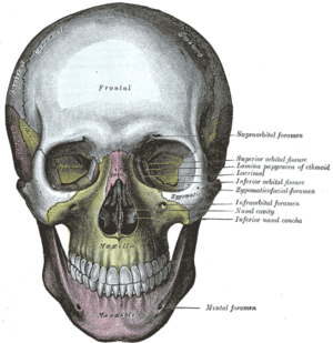
Mandible is the largest and strongest bone of the human skull. It is commonly known as the lower jaw and is located inferior to the maxilla. It is composed of a horseshoe-shaped body which lodges the teeth, and a pair of rami which projects upwards to form a temporomandibular joint.[1][2]
Structure[edit | edit source]
Mandible is formed by a body and a pair of rami.
Body[edit | edit source]
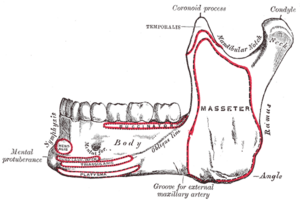
Body is the anterior portion of the mandible. Body has two surfaces: outer and inner and two borders: upper and lower border. [2]The body ends and the rami begin on either side at the angle of the mandible, also known as the gonial angle. [1]
- Outer surface is also known as external surface and has following characteristics:
- Mandibular symphysis/ Symphysis menti at midline which joins left and right half of the bone, detected as a subtle ridge in the adult.
- The inferior portion of the ridge divides and encloses a midline depression called the mental protuberance, also known as chin. The edges of the mental protuberance are elevated, forming the mental tubercle.
- Laterally to the ridge and below the incisive teeth is a depression known as the incisive fossa.
- Below the second premolar is the mental foramen, in which the mental nerve and vessels exit.
- The oblique line courses posteriorly from the mental tubercle to the anterior border of the ramus.
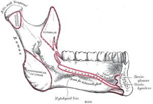
- Inner surface is also known as internal surface and has following features:
- The mylohyoid line is a prominent ridge that runs obliquely downwards and forwards from below the third molar tooth to the median area below the genial tubercles.
- Below the mylohyoid line, the surface is slightly hollowed out to form the sub-mandibular fossa, which lodges the submandibular gland.
- Above the mylohyoid line, there is the sublingual fossa in which the sublingual gland lies.
- The posterior surface of the symphysis menti is marked by four small elevations called the superior and inferior genial tubercles.
- Upper border (Alveolar border)
- It consist of sockets for the teeth.
- Lower border (Inferior border)
- It is also known as base. There is a fossa present at the side of midline known as digastric fossa. [2]
Ramus[edit | edit source]
The ramus is lateral continuation of the body and is quadrilateral in shape. The coronoid process is the anterosuperior projection of the ramus which is triangular in shape. Whereas posterosuperior projection of ramus is known as condyloid process whose head is covered with fibrocartilage and form a temporomandibular joint. The constricted part below condyloid process is neck. Condyloid and coronoid process are separated by a mandibular notch. It has two surfaces: medial and lateral and four borders: superior, inferior, anterior and posterior.[1]
Muscle Attachment[edit | edit source]
Ossification[edit | edit source]
add text here relating to diagnostic tests for the condition
[edit | edit source]
add links to outcome measures here (see Outcome Measures Database)
Clinical Relevance[edit | edit source]
add text here relating to management approaches to the condition
Differential Diagnosis
[edit | edit source]
add text here relating to the differential diagnosis of this condition
Resources
[edit | edit source]
add appropriate resources here
References[edit | edit source]
- ↑ 1.0 1.1 1.2 Breeland G, Aktar A, Patel BC. Anatomy, head and neck, mandible. StatPearls [Internet]. 2021 Jun 18.
- ↑ 2.0 2.1 2.2 BD_Chaurasia’s_Human_Anatomy, Volume 3 - Head-Neck and Brain 6th Edition. Page number:32-36
- ↑ MANDIBLE - GENERAL FEATURES & ATTACHMENTS. Available from: https://www.youtube.com/watch?v=jp55Ohh1kDY lasted accessed: 2022-02-08


