Management of Thoracic Outlet Syndrome
Medical Management[edit | edit source]
- Nonsteroidal anti-inflammatory drugs have been prescribed to reduce pain and inflammation.
- Botulinum injections to the anterior and middle scalenes have also been found to temporarily reduce pain and spasm from neurovascular compression, further research is needed because there are discrepancies in the literature[1][2].[3]
- Surgical management of TOS should only be considered after conservative treatment has been proven ineffective.[4]. However, limb-threatening complications of vascular TOS have been indicated for surgical intervention.[5]
- Surgery to treat thoracic outlet syndrome may be performed using several different approaches, including: transaxillary approach, supraclavicular approach and infraclavicular approach.[4]
Transaxillary approach.[edit | edit source]
The first rib forms the common denominator for all causes of nerve and artery compression in this region, so that its removal generally improves symptoms. The surgeon makes an incision in the chest to access the first rib, divides the muscles in front of the rib, and removes a portion of the first rib to relieve compression, without disturbing the nerves or blood vessels[6][7].[4]
Supraclavicular approach[edit | edit source]
it has been advocated to perform first rib resection and scalenectomy, a safe and effective procedure, characterized by a shorter operative time and a complication rate lower or comparable to that of transaxillary first rib resection. This approach repairs compressed blood vessels. The surgeon makes an incision just under the neck to expose the brachial plexus region. Then he looks for signs of trauma or muscles contributing to compression near the first rib. The first rib may be removed if necessary to relieve compression[7][6].[4]
Infraclavicular approach.[edit | edit source]
In this approach, the surgeon makes an incision under the collarbone and across the chest. This procedure may be used to treat compressed veins that require extensive repair.[4][7]
Neurogenic TOS[edit | edit source]
Surgical decompression should be considered for those with true neurological signs or symptoms. These include weakness, wasting of the hand intrinsic muscles, and conduction velocity less than 60 m/sec. The first rib can be a major contributor to TOS. There is controversy, however, regarding the necessity of a complete resection to reduce the chance of reattachment of the scalenes, scar tissue development, or bony growth of the remaining tissue. In addition to the first rib, cervical ribs are removed, scalenectomies can be performed, and fibrous bands can be excised[5]. Terzis found that the supraclavicular approach to treatment was an effective and precise surgical method[8]
Arterial TOS:[edit | edit source]
Decompression can include cervical and/or first rib removal and scalene muscle revision. The subclavian can then be inspected for degeneration, dilation, or aneurysm. Saphenous vein graft or synthetic prosthesis can then be used if necessary[5]
Venous TOS:[edit | edit source]
Thrombolytic therapy is the first line of treatment for these patients. Because of the risk of recurrence, many recommend that removal of the first rib is necessary even when thrombolytic therapy completely opens the vein. The results of a study show that the infraclavicular approach is a safe and effective treatment for acute VTOS. They had no brachial plexus or phrenic nerve injuries[9].[7] Angioplasty can then be used to treat those with venous stenosis[5]
In venous or arterial TOS, medication can be administered to dissolve blood clots prior to thoracic outlet compression. It may also be necessary to conduct a procedure to remove a clot from the vein or artery or repair the vein or artery prior to thoracic outlet decompression.[9]
Some larger-chested women have sagging shoulders that increase pressure on the neurovascular structures in the thoracic outlet. A supportive bra with wide and posterior-crossing straps can help reduce tension. Extreme cases may resort to breast-reduction surgery to relieve TOS and other biomechanical problems[10][11].[12]
Physical Therapy Management[edit | edit source]
Conservative management should be the first strategy to treat TOS since this seems to be effective at decreasing symptoms, facilitating return to work, and improving function, but yet a few studies have evaluated the optimal exercise program as well as the difference between conservative management and no treatment. [13]
Conservative management includes physical therapy, which focuses mainly on patient education, pain control, range of motion, nerve gliding techniques, strengthening and stretching[14].
Stage 1[edit | edit source]
The aim of the initial stage is to decrease the patient’s symptoms. This may be achieved by patient education, in which TOS, bad postures, the prognosis and the importance of therapy compliance are explained. Furthermore, some patients who sleep with the arms in an overhead, abducted position should get some information about their sleeping posture to avoid waking up at night. These patients should sleep on their uninvolved side or supine, potentially by pinning down the sleeves. The Cyriax release test may be used if a ‘release phenomenon’ is present. This technique completely unloads the neurovascular structures in the thoracic outlet before going to bed.[5]
Cyriax Release Maneuver[edit | edit source]
- Elbows flexed to 90°
- Towels create a passive shoulder girdle elevation
- Supported spine and the head in neutral
- The position is held until peripheral symptoms are produced. The patient is encouraged to allow symptoms to occur as long as can be tolerated for up to 30 minutes, observing for a symptom decrescendo as time passes.[5]
The patient’s breathing techniques need to be evaluated as the scalenes and other accessory muscles often compensate to elevate the ribcage during inspiration. Encouraging diaphragmatic breathing will lessen the workload on already overused or tight scalenes and can possibly reduce symptoms.[5]
Scapula Settings and Control[edit | edit source]
In the treatment, you first have to start with scapula settings and control.
This is important to establishing normal scapula muscle recruitment and control in the resting position. Once this is achieved then the program is progressed to maintaining scapula control while both motion and load are applied. The programme begins in lower ranges of abduction and is gradually progressed further up into abduction and flexion range until muscles are being retrained in functional movement patterns at higher ranges of elevation.
Control the Humeral Head Position[edit | edit source]
It is also important to control the humeral head position. Specific drills are given to facilitate humeral head control. The most common aberrant position of the humeral head is an increase in anterior placement of the humeral head. A useful strategy to help facilitate co-contraction of the rotator cuff to help stabilize and centralize the humeral head is to facilitate a mid-level isometric contraction of the rotator cuff by applying resistance to the humeral head (Dark et al., 2007).
Further on in the treatment, this may be integrated into movement patterns. First in slow controlled concentric/eccentric motion drills, later isolated muscle strengthening drills.
Serratus Anterior Recruitment and Control[edit | edit source]
Abduction external rotation strategies described above are often sufficient to trigger serratus anterior recruitment and control without the risk of over-activating pectoral minor muscle
Stage 2:[edit | edit source]
Once the patient has control over his/her symptoms, the patient can move to this stage of treatment. The goal of this stage is to directly address the tissues that create structural limitations of motion and compression. How this should be done is one of the most discussed topics of this pathology. Some examples of methods that are used in the literature are.
- Massage
- Strengthening of the levator scapulae, sternocleidomastoid and upper trapezius (This group of muscles open the thoracic outlet by raising the shoulder girdle and opening the costoclavicular space)
- Stretching of the pectoralis, lower trapezius and scalene muscles (These muscles close the thoracic outlet)
- Postural correction exercises
- Relaxation of shortened muscles[10]
- Aerobic exercises in a daily home exercise program[15][10]
Exercises[edit | edit source]
- Shoulder exercises to restore the range of motion and so provide more space for the neurovascular structures. Exercise: Lift your shoulders backwards and up, flex your upper thoracic spine and move the shoulders forward and down. Then straighten the back and repeat 5 to 10 times.
- ROM of the upper cervical spine. Exercise: Lower your chin 5 to 10 times against your chest, while you are standing with the back of your head against a wall. The effectiveness of this exercise can be enlarged by pressing the head down by hands.
- Activation of the scalene muscles is the most important exercises. These exercises help to normalize the function of the thoracic aperture as well as all the malfunctions of the first rib. Exercises are Anterior scalene (Press your forehead 5 times against the palm of your hand for a duration of 5 seconds, without creating any movement), Middle scalene (Press your head sidewards against your palm), Posterior scalene (Press your head backwards against your palm
- Stretching exercises
Other Interventions[edit | edit source]
- Repositioning/mobilization of the shoulder girdle and pelvis joints: cervicothoracic, sternoclavicular, acromioclavicular, and costotransverse joints[5] [10]
- Glenohumeral mobilizations in end-range elevation with the elbow supported in extension[15]
- Taping: some patients with severe symptoms respond to additional taping, adhesive bandages or braces that elevate or retract the shoulder girdle.[16][13]
Manipulative Treatment to Mobilize the First Rib[edit | edit source]
These should be carried out with caution and only after a thorough assessment as they can provoke irritation and pain symptoms in some patients[16]
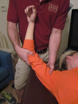
- Posterior Glenohumeral Glide with Arm Flexion: The patient is supine. The mobilization hand contacts the proximal humerus avoiding corocoid process. The force is directed posterolaterally (direction of thumb).
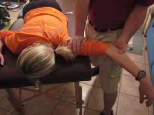
- Anterior Glenohumeral Glide with Arm Scaption: The patient is prone. The mobilization hand contacts the proximal humerus avoiding acromion process. The force is directed anteromedially.
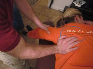
- Inferior Glenohumeral Glide: The patient is prone. The stabilizing hand holds the proximal humerus, the humerus distal to the lateral acromion process. The mobilization hand contacts the axillary border of the scapula. Mobilize the scapula in a craniomedial direction along the ribcage.[5]
First Rib Mobilization: Patient seated. Thin sheet strap positioned around the first rib. Pull strap towards the opposite hip. Neck retracted, contralateral lateral flexion, and ipsilateral rotation. Ipsilateral head rotation emphasizes scalene stretch. Contralateral rotation emphasizes rib mobilization.
Posterior Glenohumeral Glide with Arm Flexion: Patient supine. Mobilizing hand contacts proximal humerus avoiding corocoid process. Force is directed posterolaterally (direction of thumb).
Anterior Glenohumeral Glide with Arm Scaption: Patient prone. Mobilizing hand contacts proximal humerus avoiding acromion process. Force is anteromedially.
Inferior Glenohumeral Glide: Patient prone. Stabilizing hand holds proximal humerus. Mobilizing hand contacts axillary border of scapula. Mobilize scapula in craniomedial direction along ribcage.
Post-Op Physical Therapy[edit | edit source]
If a patient does require surgery, then physical therapy should follow immediately to prevent scar tissue and return the patient to full function.
- ↑ Jordan SE, Ahn SS, Freischlag JA, Gelabert HA, Machleder HI. Selective botulinum chemodenervation of the scalene muscles for treatment of neurogenic thoracic outlet syndrome. Annals of vascular surgery. 2000 Jul 1;14(4):365-9.
- ↑ Christo PJ, Christo DK, Carinci AJ, Freischlag JA. Single CT-guided chemodenervation of the anterior scalene muscle with botulinum toxin for neurogenic thoracic outlet syndrome. Pain Medicine. 2010 Apr 1;11(4):504-11.
- ↑ Finlayson HC, O’Connor RJ, Brasher PM, Travlos A. Botulinum toxin injection for management of thoracic outlet syndrome: a double-blind, randomized, controlled trial. Pain. 2011 Sep 1;152(9):2023-8.
- ↑ 4.0 4.1 4.2 4.3 4.4 Ciampi P, Scotti C, Gerevini S, De Cobelli F, Chiesa R, Fraschini G, Peretti GM. Surgical treatment of thoracic outlet syndrome in young adults: single centre experience with minimum three-year follow-up. International orthopaedics. 2011 Aug;35(8):1179-86.
- ↑ 5.0 5.1 5.2 5.3 5.4 5.5 5.6 5.7 5.8 Hooper TL, Denton J, McGalliard MK, Brismée JM, Sizer Jr PS. Thoracic outlet syndrome: a controversial clinical condition. Part 2: non-surgical and surgical management. Journal of Manual & Manipulative Therapy. 2010 Sep 1;18(3):132-8.
- ↑ 6.0 6.1 Urschel Jr HC, Razzuk MA. Neurovascular compression in the thoracic outlet: changing management over 50 years. Annals of surgery. 1998 Oct;228(4):609
- ↑ 7.0 7.1 7.2 7.3 Siracuse JJ, Johnston PC, Jones DW, Gill HL, Connolly PH, Meltzer AJ, Schneider DB. Infraclavicular first rib resection for the treatment of acute venous thoracic outlet syndrome. Journal of Vascular Surgery: Venous and Lymphatic Disorders. 2015 Oct 1;3(4):397-400.
- ↑ Terzis JK, Kokkalis ZT. Supraclavicular approach for thoracic outlet syndrome. Hand. 2010 Sep 1;5(3):326-37.
- ↑ 9.0 9.1 Schneider DB, Dimuzio PJ, Martin ND, Gordon RL, Wilson MW, Laberge JM, Kerlan RK, Eichler CM, Messina LM. Combination treatment of venous thoracic outlet syndrome: open surgical decompression and intraoperative angioplasty. Journal of vascular surgery. 2004 Oct 1;40(4):599-603.
- ↑ 10.0 10.1 10.2 10.3 Vanti C, Natalini L, Romeo A, Tosarelli D, Pillastrini P. Conservative treatment of thoracic outlet syndrome. Eura medicophys. 2007 Mar 1;43:55-70.
- ↑ Buckley L, Schub E. Thoracic Outlet Syndrome. October 2010; Accessed November 2,2011
- ↑ Boezaart AP, Haller A, Laduzenski S, Koyyalamudi VB, Ihnatsenka B, Wright T. Neurogenic thoracic outlet syndrome: A case report and review of the literature. International journal of shoulder surgery. 2010 Apr;4(2):27.
- ↑ 13.0 13.1 Vanti C. et al., Conservative treatment of thoracic outlet syndrome A review of the literature, Europa Medicophysica, 2006, Volume 42
- ↑ Crosby CA, Wehbé MA. Conservative treatment for thoracic outlet syndrome. Hand clinics. 2004 Feb 1;20(1):43-9.
- ↑ 15.0 15.1 Lindgren KA. Conservative treatment of thoracic outlet syndrome: a 2-year follow-up. Archives of physical medicine and rehabilitation. 1997 Apr 1;78(4):373-8.
- ↑ 16.0 16.1 Vanti C, Natalini L, Romeo A, Tosarelli D, Pillastrini P. Conservative treatment of thoracic outlet syndrome. Eura medicophys. 2007 Mar 1;43:55-70.


