Lumbar Stress Fractures in Cricket Players: Difference between revisions
(Added movement available and pathology) |
(moved images around) |
||
| Line 1: | Line 1: | ||
'''Relevant Anatomy''' | '''<big>Relevant Anatomy</big>''' | ||
[[File:Lumbar joints image.png|thumb|There are three joints between lumbar vertebrae two facet joints (zygapophyseal joints), one at either side and a central vertebral disc joint.]] | [[File:Lumbar joints image.png|thumb|There are three joints between lumbar vertebrae two facet joints (zygapophyseal joints), one at either side and a central vertebral disc joint.]] | ||
The lumbar vertebrae is made up of 5 different bones L1 is the most superior and L5 the most inferior. These 5 vertebrae share most of the same features and have very similar function. All stress fractures in the lumbar spine occur at either L3 (12% of stress fractures), L4 (35% of stress fractures) or L5 (32% of stress fractures). The remaining percentages account for fractures in multiple locations (Alway et al, 2019). | The lumbar vertebrae is made up of 5 different bones L1 is the most superior and L5 the most inferior. These 5 vertebrae share most of the same features and have very similar function. All stress fractures in the lumbar spine occur at either L3 (12% of stress fractures), L4 (35% of stress fractures) or L5 (32% of stress fractures). The remaining percentages account for fractures in multiple locations (Alway et al, 2019). | ||
There are three joints between each vertebra, two facet joints (also known as zygapophyseal joints) which are on either side and also a vertebral disc joint in the centre of the vertebrae. These joints do not allow for much movement as the lumbar structures are very stable. | There are three joints between each vertebra, two facet joints (also known as zygapophyseal joints) which are on either side and also a vertebral disc joint in the centre of the vertebrae. These joints do not allow for much movement as the lumbar structures are very stable. | ||
''' | '''Movement at the lumbar joints''' | ||
Different joints in the lumbar spine allow for differing degrees of movement. All of the lumbar spine joints allow for most movement within the sagittal plane allowing for flexion and extension. L1 allows for around 12 degrees whereas L5 allows for around 17 degrees (these numbers change based on individual differences). | |||
This is why lumbar stress fractures occur at the lower vertebrae as lumbar stress fractures occur as a result of extension related forces so the vertebrae that allow for more extension are more likely to be injured. | |||
[[File:Chart of movement in lumbar spine.png|thumb|856x856px|The movement available at different joints in the lumbar spine and the degrees of movement that each movement can reach at the joint. '''Yellow''' Flexion/Extension '''Blue''' Rotation '''Purple''' Side flexion |center]] | |||
[[File:Labelled lumbar vertebrae .png|thumb|This is a typical lumbar vertebrae, note the pedicle labelled on the left of the image 22.8% of lumbar stress fractures occur here.]] | '''<big>Stress fractures</big>''' | ||
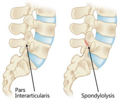
[[File:Pars Interarticularis and spondylolysis.png|thumb|77.2% of lumbar stress fractures occur at the Pars Interarticularis, these fractures are also known as spondylolysis'.|244x244px]] | |||
[[File:Labelled lumbar vertebrae .png|thumb|This is a typical lumbar vertebrae, note the pedicle labelled on the left of the image 22.8% of lumbar stress fractures occur here.|256x256px]] | |||
Stress fractures are small cracks within a bone. For something to be defined as a stress fracture there must be a clear crack in the bone without it being a complete crack as that would be a different type of fracture(Astur et al., 2016). | |||
In the lumbar spine this crack can appear in two different locations. The pars interarticularis (77.2% of lumbar stress fractures) and the pedicle (22.8% of stress fractures) (Alway et al, 2019). A stress fracture at the pars interarticularis is more frequently referred to as a spondylolysis. | |||
'''<big>Pathology</big> ''' | |||
Stress fractures are overuse injuries. In the presence of repetitive loading and inadequate time for healing and recovery. The repetitive stress response leads to increased osteoclastic activity surpassing the rate of osteoblastic activity, this causes weakening of the bone making it susceptible to stress fracture. Over time the fracture develops. Fatigue in other structures around the vertebrae is a common cause of the stress fracture. | Stress fractures are overuse injuries. In the presence of repetitive loading and inadequate time for healing and recovery. The repetitive stress response leads to increased osteoclastic activity surpassing the rate of osteoblastic activity, this causes weakening of the bone making it susceptible to stress fracture. Over time the fracture develops. Fatigue in other structures around the vertebrae is a common cause of the stress fracture. | ||
Revision as of 21:09, 18 March 2024
Relevant Anatomy
The lumbar vertebrae is made up of 5 different bones L1 is the most superior and L5 the most inferior. These 5 vertebrae share most of the same features and have very similar function. All stress fractures in the lumbar spine occur at either L3 (12% of stress fractures), L4 (35% of stress fractures) or L5 (32% of stress fractures). The remaining percentages account for fractures in multiple locations (Alway et al, 2019).
There are three joints between each vertebra, two facet joints (also known as zygapophyseal joints) which are on either side and also a vertebral disc joint in the centre of the vertebrae. These joints do not allow for much movement as the lumbar structures are very stable.
Movement at the lumbar joints
Different joints in the lumbar spine allow for differing degrees of movement. All of the lumbar spine joints allow for most movement within the sagittal plane allowing for flexion and extension. L1 allows for around 12 degrees whereas L5 allows for around 17 degrees (these numbers change based on individual differences).
This is why lumbar stress fractures occur at the lower vertebrae as lumbar stress fractures occur as a result of extension related forces so the vertebrae that allow for more extension are more likely to be injured.
Stress fractures
Stress fractures are small cracks within a bone. For something to be defined as a stress fracture there must be a clear crack in the bone without it being a complete crack as that would be a different type of fracture(Astur et al., 2016).
In the lumbar spine this crack can appear in two different locations. The pars interarticularis (77.2% of lumbar stress fractures) and the pedicle (22.8% of stress fractures) (Alway et al, 2019). A stress fracture at the pars interarticularis is more frequently referred to as a spondylolysis.
Pathology
Stress fractures are overuse injuries. In the presence of repetitive loading and inadequate time for healing and recovery. The repetitive stress response leads to increased osteoclastic activity surpassing the rate of osteoblastic activity, this causes weakening of the bone making it susceptible to stress fracture. Over time the fracture develops. Fatigue in other structures around the vertebrae is a common cause of the stress fracture.
In healthy bone repair occurs thanks to osteoblastic activity. However, when the recovery period is insufficient for long periods of time the boney repair doesn’t happen and so the repetitive loading causes microfractures which develop into full stress fractures(Kiel and Kaiser, 2020).
Biomechanical Underpinnings The fast-bowling action is a complex sequence of movements that places significant stress on the lumbar spine. The action commences during the delivery stride prior to ball release, with the first key event being back foot contact (BFC) when the bowler's back foot impacts the ground (Bartlett, 1996). This is followed by front-foot contact (FFC) after ball release. Fast bowling actions can be broadly categorised into one of four types: front-on, side-on, semi-open, and mixed (Thiagarajan et al., 2015). This categorisation is determined according to the alignment of the shoulders at BFC and the amount of shoulder counter-rotation during the delivery stride (Portus et al., 2004).
[Image 1: Illustration of the four bowling action types]
The Mixed Bowling Action Research by Cook et al. (2015) identifies a strong correlation between the "mixed action" bowling style and an increased prevalence of lumbar stress fractures. This action is characterised by a significant angular disparity (>40 degrees) between the hips and shoulders, as well as excessive counter-rotation of the torso. This action leads to several detrimental effects: Excessive extension and rotation, placing undue stress on the pars interarticularis, a vulnerable region of the vertebrae. Repetitive lumbar hyperextension, with the bowler's front foot landing while the torso is in a hyperextended position. This places high loads on the posterior elements of the lumbar vertebrae, particularly the pars interarticularis, leading to potential microfractures over time.
Rotational forces, as the bowling action involves a forceful rotation of the torso to generate pace. This rotation, combined with hyperextension, creates a complex stress pattern on the spine, further increasing the risk of fractures. Lateral flexion, where some bowlers, particularly younger athletes, may exhibit uncontrolled lateral flexion of the spine during the delivery stride. This uneven loading on the vertebrae increases the risk of injury, as evidenced by Hibbert et al. (2018). Their research introduced the "crunch factor," which considers both lateral flexion and rotational velocity of the lumbar spine. A higher "crunch factor" has been linked to a greater risk of stress fractures on the side opposite the bowling arm.
[Image 2: Illustration of the mixed bowling action and associated forces]
Ground Reaction Forces and Altered Stress Distribution During the fast bowling delivery stride, peak ground reaction forces can reach up to nine times the bowler's body weight (Linthorne et al., 2015). These substantial forces are transmitted through the body, impacting the lumbar facet joints. If the bowler's front foot lands while the spine is in a combined state of rotation, extension, and lateral flexion, the stress distribution on the spine is disrupted (Linthorne et al., 2015). This abnormal loading places excessive strain on the pars interarticularis, as the forces are not absorbed efficiently, increasing the fracture risk.
[Image 3: Diagram showing ground reaction forces and stress distribution]
Bowling Workload and Fatigue-Induced Technique Breakdown First-class bowlers can deliver a significant number of balls (300-500) per week during a season (Linthorne et al., 2015). Inadequate workload management can have detrimental consequences. Research by Gabbett et al. (2011) suggests that bowling too many overs in a single session can lead to fatigue, causing technique to deteriorate. This can manifest as increased counter-rotation, further stressing the lumbar spine. Additionally, a study by Cumming et al. (2015) demonstrated that junior fast bowlers with less than 3.5 days of recovery between bowling sessions were three times more likely to sustain injuries, including lumbar stress fractures.
Risk Factors Beyond Biomechanics While biomechanics play a central role, several other factors contribute to a fast bowler's susceptibility to lumbar stress fractures. Improper bowling mechanics, such as excessive shoulder counter-rotation in adolescents or uncontrolled trunk rotation in adults, can place additional stress on the lumbar spine. Fast bowlers with high delivery volumes, especially those experiencing rapid increases in workload (e.g., transitioning from junior to senior cricket), are more likely to develop stress fractures due to insufficient time for bone adaptation.
Adolescent bowlers (aged 15-18) are particularly vulnerable due to their growing skeletons. Their bones are not yet fully mature and can be less tolerant of the repetitive stress of fast bowling. A weak core or imbalances between core muscle groups can lead to uneven stress distribution on the spine, compromising its stability and increasing the risk of fractures. Bowlers with pre-existing lower back issues are also at greater risk. Furthermore, bowlers with larger body sizes may experience higher loads on their lumbar spine during bowling, and inadequate levels of vitamin D and bone calcium can compromise bone health and increase susceptibility to stress fractures.
Lumbar stress fractures are a significant concern for fast bowlers in cricket, with the biomechanics of the bowling action and various risk factors contributing to their development. By understanding the underlying mechanisms, such as the impact of mixed bowling actions, ground reaction forces, and workload management, teams can implement targeted preventive measures. These measures include technical coaching, workload monitoring, strength and conditioning programs, and early screening and intervention. Through a comprehensive approach, cricket teams can mitigate the risk of these debilitating injuries, ensuring the longevity and success of their fast bowling talents.


