Iliotibial Tract
Top Contributors - Eman Ammar and Lucinda hampton
Description[edit | edit source]
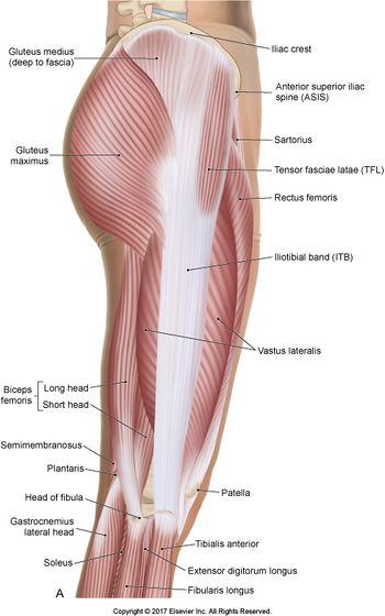
The iliotibial tract or iliotibial band is a longitudinal fibrous reinforcement of the fascia lata.runs along the lateral thigh and serves as an important structure involved in lower extremity motion.
The part of the iliotibial band which lies beneath the tensor fasciae latae is prolonged upward to join the lateral part of the capsule of the hip-joint. The tensor fasciae latae effectively tightens the iliotibial band around the area of the knee. This allows for bracing of the knee especially in lifting the opposite foot.
The gluteus maximus muscle and the tensor fasciae latae insert upon the tract.
Origin[edit | edit source]
It originates at the anterolateral iliac tubercle portion of the external lip of the iliac crest
Insertion[edit | edit source]
inserts at the lateral condyle of the tibia at Gerdy's tubercle
Nerve[edit | edit source]
the ITB shares the innervation of the TFL and gluteus maximus via the superior gluteal nerve (SGN) and inferior gluteal nerve (IGN)
Artery[edit | edit source]
The ITB, being a tendinous extension of the tensor fascia lata (TFL), shares the same arterial supply:
- Ascending branch of the lateral femoral circumflex artery (LFCA)
- Superior gluteal artery (SGA)
Function.[edit | edit source]
The action of the muscles associated with the ITB (TFL and some fibers of Gluteus Maximus) flex, extend, abduct, and laterally and medially rotate the hip. The ITB contributes to lateral knee stabilization. During knee extension the ITB moves anterior to the lateral condyle of the femur, while ~30 degrees knee flexion, the ITB moves posterior to the lateral condyle. However, it has been suggested that this is only an illusion due to the changing tension in the anterior and posterior fibers during movement
Clinical relevance[edit | edit source]
External snapping hip syndrome, or externa coxa saltans has the potential to cause chronic pain in the lateral aspect of the hip located over the greater trochanter of the femur. Pathophysiology comprises thickening of the posterior aspect of the ITB or anterior tendon fibers of the gluteus maximus muscle near its insertion. This portion of the band remains posterior to the greater trochanter in hip extension, however, moves anteriorly when flexed, adducted, or internally rotated causing a "snapping" mechanism. This snapping is the tense fascial structure catching on the greater trochanter as it moves in the before mentioned motions.
Assessment[edit | edit source]
Clinical examination testing for ITB dysfunction is best elicited utilizing the Ober Test.
To perform the Ober test, have the patient lie on his or her uninvolved side in the lateral decubitus position. The symptomatic side should be facing upward (i.e., closest to the ceiling). Next, the examiner passively flexes the knee to about 90 degrees. The hip is then brought passively into a flexed and abducted position. Next, the examiner assesses the passive flexibility over the ITB with the hip brought into increasing levels of extension and adduction. A positive test entails pain, tightness, or clicking over the ITB.
Treatment[edit | edit source]
Treatment generally initiates with ITB stretching and physical therapy. NSAID use may be beneficial to reduce inflammation. Surgery is a last resort used for refractory cases
Resources[edit | edit source]
- ↑ Evans P. The postural function of the iliotibial tract. Ann R Coll Surg Engl. 1979 Jul;61(4):271-80. [PMC free article] [PubMed] 2. Strauss EJ, Kim S, Calcei JG, Park D. Iliotibial band syndrome: evaluation and management. J Am Acad Orthop Surg. 2011 Dec;19(12):728-36. [PubMed] 3. Chahla J, Murray IR, Robinson J, Lagae K, Margheritini F, Fritsch B, Leyes M, Barenius B, Pujol N, Engebretsen L, Lind M, Cohen M, Maestu R, Getgood A, Ferrer G, Villascusa S, Uchida S, Levy BA, Von Bormann R, Brown C, Menetrey J, Hantes M, Lording T, Samuelsson K, Frosch KH, Monllau JC, Parker D, LaPrade RF, Gelber PE. Posterolateral corner of the knee: an expert consensus statement on diagnosis, classification, treatment, and rehabilitation. Knee Surg Sports Traumatol Arthrosc. 2019 Aug;27(8):2520-2529. [PubMed] 4. Musick SR, Varacallo M. StatPearls [Internet]. StatPearls Publishing; Treasure Island (FL): Apr 28, 2020. Snapping Hip Syndrome. [PubMed]


