Flexor retinaculum: Difference between revisions
Leana Louw (talk | contribs) No edit summary |
No edit summary |
||
| Line 6: | Line 6: | ||
== Description == | == Description == | ||
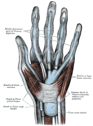
[[File:Flexor retinaculum of hand.png|alt=Flexor retinaculum of hand.png |thumb|'''Flexor retinaculum of hand''']] | [[File:Flexor retinaculum of hand.png|alt=Flexor retinaculum of hand.png |thumb|'''Flexor retinaculum of hand''']] | ||
Flexor retinaculum is a strong fibrous band which bridges the anterior concavity of the carpal bones thus converts it into a tunnel, the carpal tunnel<ref name=":0"> | Flexor retinaculum is a strong fibrous band which bridges the anterior concavity of the carpal bones thus converts it into a tunnel, the carpal tunnel<ref name=":0">Chaurasia BD.Human Anatomy.Vol.1.Sixth Edition.</ref>. | ||
=== Attachments === | === Attachments === | ||
| Line 19: | Line 19: | ||
On either side the retinaculum has a slip. | On either side the retinaculum has a slip. | ||
* ''Lateral deep slip'' - It is attached to the medial lip of the groove on the trapezium thus converts it into a fibro-osseous tunnel that transmits the tendon of the flexor carpi radialis and its synovial sheath. | * ''Lateral deep slip'' - It is attached to the medial lip of the groove on the trapezium thus converts it into a fibro-osseous tunnel that transmits the tendon of the flexor carpi radialis and its synovial sheath. | ||
* ''Medial superficial slip'' - It is attached to the pisiform bone and it forms a small canal (of Guyon). The ulnar vessels and nerves pass deep to this slip. Occasionally Compression of the [[Ulnar Nerve|ulnar nerve]] may occur within this canal. <ref name=":0" /> <ref> | * ''Medial superficial slip'' - It is attached to the pisiform bone and it forms a small canal (of Guyon). The ulnar vessels and nerves pass deep to this slip. Occasionally Compression of the [[Ulnar Nerve|ulnar nerve]] may occur within this canal. <ref name=":0" /> <ref>McMinn RMH, Last's Anatomy Regional and Applied,Ninth edition</ref> | ||
{{#ev:youtube|J47tdKW3ibg|250}} <ref>Dr | {{#ev:youtube|J47tdKW3ibg|250}} <ref>Dr.Prakash GB. The flexor retinaculum of Hand : Gross anatomy, attachments and relations. Available from:https://www.youtube.com/watch?v=J47tdKW3ibg&feature=youtu.be | ||
</ref> | </ref> | ||
Revision as of 20:11, 8 October 2020
Original Editor - Shanshika Maddumage
Top Contributors - Shanshika Maddumage and Leana Louw
Description[edit | edit source]
Flexor retinaculum is a strong fibrous band which bridges the anterior concavity of the carpal bones thus converts it into a tunnel, the carpal tunnel[1].
Attachments[edit | edit source]
Medially,
- To the pisiform bone
- To the hook of the hamate
Laterally,
These four bony points are all palpable in the living hand and it should be noted that pisiform is the only carpal bone that gives attachments to both flexor and extensor retinacula.
On either side the retinaculum has a slip.
- Lateral deep slip - It is attached to the medial lip of the groove on the trapezium thus converts it into a fibro-osseous tunnel that transmits the tendon of the flexor carpi radialis and its synovial sheath.
- Medial superficial slip - It is attached to the pisiform bone and it forms a small canal (of Guyon). The ulnar vessels and nerves pass deep to this slip. Occasionally Compression of the ulnar nerve may occur within this canal. [1] [2]
Function[edit | edit source]
Principal function of the flexor retinaculum is to serve as a pulley for the carpal flexor muscles and to stabilize the carpal system [4].
In addition,
- The volar surface gives rise to muscles of the thenar and hypothenar eminences
- It is Related to the tendon of the palmaris longus
- Its Upper margin continues in the palmar carpal ligament and lower margin merges with the palmar aponeurosis
Clinical relevance[edit | edit source]
Carpal tunnel syndrome results when the retinaculum compresses the underlying median nerve[4]
References[edit | edit source]
- ↑ 1.0 1.1 1.2 Chaurasia BD.Human Anatomy.Vol.1.Sixth Edition.
- ↑ McMinn RMH, Last's Anatomy Regional and Applied,Ninth edition
- ↑ Dr.Prakash GB. The flexor retinaculum of Hand : Gross anatomy, attachments and relations. Available from:https://www.youtube.com/watch?v=J47tdKW3ibg&feature=youtu.be
- ↑ 4.0 4.1 Deak N, Bordoni B. Anatomy, Shoulder and Upper Limb, Wrist Flexor Retinaculum.


