File:HP AP weight bearing.png: Difference between revisions
(Diagram of the pelvis in the frontal plane (anterior view) depicting arrows where the socket fits tightly over the iliac crests and between the greater trochanter to prevent shifting and pistoning inside the socket when walking.) |
No edit summary |
||
| Line 1: | Line 1: | ||
== Summary == | == Summary == | ||
Hemipelvectomy prosthetic socket fitting: this diagram of the pelvis in the frontal plane (anterior view) depicting arrows where the socket fits tightly over the iliac crests and between the greater trochanter to prevent shifting and pistoning inside the socket when walking. | |||
== Licensing == | == Licensing == | ||
{{allow-physiopedia}} | {{allow-physiopedia}} | ||
Latest revision as of 16:07, 10 July 2022
Summary[edit | edit source]
Hemipelvectomy prosthetic socket fitting: this diagram of the pelvis in the frontal plane (anterior view) depicting arrows where the socket fits tightly over the iliac crests and between the greater trochanter to prevent shifting and pistoning inside the socket when walking.
Licensing[edit | edit source]
The copyright holder has given special permission to use this work in Physiopedia.
File history
Click on a date/time to view the file as it appeared at that time.
| Date/Time | Thumbnail | Dimensions | User | Comment | |
|---|---|---|---|---|---|
| current | 16:06, 10 July 2022 | 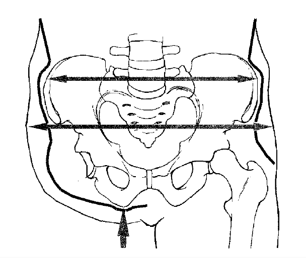 | 608 × 514 (39 KB) | Martina Lukin (talk | contribs) | Diagram of the pelvis in the frontal plane (anterior view) depicting arrows where the socket fits tightly over the iliac crests and between the greater trochanter to prevent shifting and pistoning inside the socket when walking. |
You cannot overwrite this file.
File usage
There are no pages that use this file.

