File:Diverticula.jpg
Original file (1,024 × 678 pixels, file size: 100 KB, MIME type: image/jpeg)
Summary[edit | edit source]
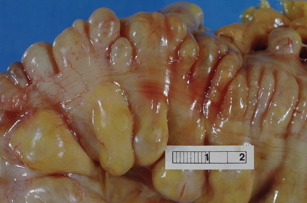
Large bowel (sigmoid colon) showing multiple diverticula. Note how the diverticula appear on either side of the longitudinal muscle bundle (taenium).
Haymanj - Self-photographed
Licensing[edit | edit source]
This work has been released into the public domain by its author. This applies worldwide. In some countries this may not be legally possible; if so: the author grants anyone the right to use this work for any purpose, without any conditions, unless such conditions are required by law.
Summary[edit | edit source]
Description: image of a vertebra Source: Modified Illustration from Anatomy & Physiology, Connexions Web site. http://cnx.org/content/col11496/1.6/
Licensing[edit | edit source]
Please include the following information in the summary box:
Image courtesy of Dr [name of contributor], Radiopaedia.org; rID:xxxx (which can be found top right in the "case information" box on case pages).
File history
Click on a date/time to view the file as it appeared at that time.
| Date/Time | Thumbnail | Dimensions | User | Comment | |
|---|---|---|---|---|---|
| current | 03:32, 6 April 2016 | | 1,024 × 678 (100 KB) | Katie Countryman (talk | contribs) | Large bowel (sigmoid colon) showing multiple diverticula. Note how the diverticula appear on either side of the longitudinal muscle bundle (taenium). Haymanj - Self-photographed |
You cannot overwrite this file.
File usage
The following page uses this file:

