File:Diverticula.jpg
Original file (1,024 × 678 pixels, file size: 100 KB, MIME type: image/jpeg)
Summary[edit | edit source]
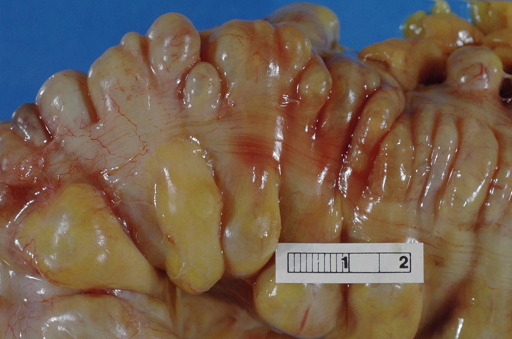
Large bowel (sigmoid colon) showing multiple diverticula. Note how the diverticula appear on either side of the longitudinal muscle bundle (taenium).
Haymanj - Self-photographed
Licensing[edit | edit source]
This work has been released into the public domain by its author. This applies worldwide. In some countries this may not be legally possible; if so: the author grants anyone the right to use this work for any purpose, without any conditions, unless such conditions are required by law.
Summary[edit | edit source]
Description: image of a vertebra Source: Modified Illustration from Anatomy & Physiology, Connexions Web site. http://cnx.org/content/col11496/1.6/
Licensing[edit | edit source]
This file is licensed under the Creative Commons Attribution-Share Alike 3.0 Unported license.
You are free:
- to share
- to copy, distribute and transmit the work to remix
- to adapt the work
Under the following conditions:
- attribution – You must attribute the work in the manner specified by the author or licensor (but not in any way that suggests that they endorse you or your use of the work).
- share alike – If you alter, transform, or build upon this work, you may distribute the resulting work only under the same or similar license to this one.
File history
Click on a date/time to view the file as it appeared at that time.
| Date/Time | Thumbnail | Dimensions | User | Comment | |
|---|---|---|---|---|---|
| current | 03:32, 6 April 2016 | | 1,024 × 678 (100 KB) | Katie Countryman (talk | contribs) | Large bowel (sigmoid colon) showing multiple diverticula. Note how the diverticula appear on either side of the longitudinal muscle bundle (taenium). Haymanj - Self-photographed |
You cannot overwrite this file.
File usage
The following page uses this file:

