Colorectal Cancer: Difference between revisions
No edit summary |
No edit summary |
||
| Line 73: | Line 73: | ||
<u>'''Radiation Therapy'''</u>''': '''helps to destroy cancer cells and can be used in conjunction with chemotherapy. <br>'''Radiation options:<br>'''Intensity Modulated Radiation Therapy (IMRT)<br>Intraoperative Radiation Therapy (IORT)<br>CyberKnife®<br>TheraShpere®<br>TomoTherapy®<br>Trilogy™<br> | <u>'''Radiation Therapy'''</u>''': '''helps to destroy cancer cells and can be used in conjunction with chemotherapy. <br>'''Radiation options:<br>'''Intensity Modulated Radiation Therapy (IMRT)<br>Intraoperative Radiation Therapy (IORT)<br>CyberKnife®<br>TheraShpere®<br>TomoTherapy®<br>Trilogy™<br> | ||
== Diagnostic Tests/Lab Tests/Lab Values == | == Diagnostic Tests/Lab Tests/Lab Values<ref name="ACS" /> == | ||
'''Preventative Scopes'''<br> | '''Preventative Scopes'''<br> | ||
Revision as of 20:16, 14 February 2013
Original Editors - Jacqueline Lopez & Abby Schnur from Bellarmine University's Pathophysiology of Complex Patient Problems project.
Lead Editors - Your name will be added here if you are a lead editor on this page. Read more.
Definition/Description[edit | edit source]
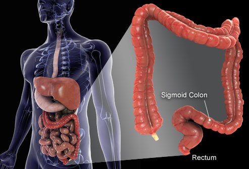
Colorectal cancer (CRC) is a rapid abnormal cell growth that affects the large intestines and/or rectum. These clusters of cells are called adenomatous polyps and develop from the tissue membrane of glandular tissue. Polyps can start as benign and non-cancerous but with time can develop and become cancerous.
Prevalence
[edit | edit source]
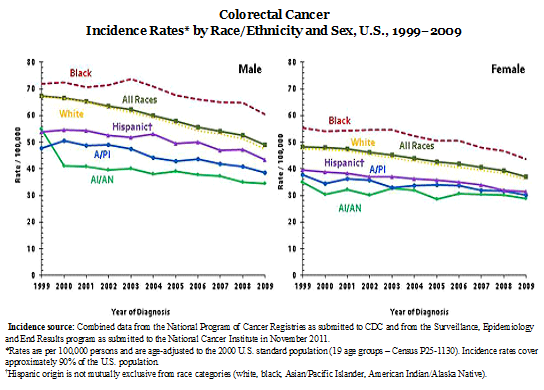
Colorectal cancer is the second leading cause of death from a type of cancer in the United States. It is also the third most common cancer among men and women. The most current statistics report 136,717 people were diagnosed in 2009 with colorectal cancer (51.26% male and 48.63% female) and 51,848 deaths (51.7% male and 48.3% female) according to the Center of Disease Control.
Data from 2009 provided by the National Cancer Institute showed that prior to January 1, 2009 1,140,161 people were living with a diagnosis of CRC in the United States. This number includes people both, currently seeking treatment for their active diagnosis, as well as, individuals who have been in years of remission. 558,648 of these individuals were male and 581,477 were female.
Other statistical facts gathered from various sources include the following:
- The lifetime risk of developing CRC is 1/20 or 4.96%.[1]
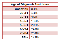
- The mean age of CRC diagnosis is 69 years of age. [2]
- The mean mortality age of CRC is 74 years of age. [2]
- Studies between 1991 and 2005 show that survival rates from CRC have increased by 30%. [3]
- The risk of getting CRC increases with age and is greater in men than women. [4]
- The most common area of diagnosis is the rectum and the rectosigmoid junction, with the sigmoid resulting the most favorable outcome. [5]
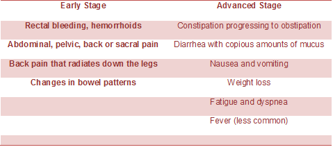
Characteristics/Clinical Presentation[edit | edit source]
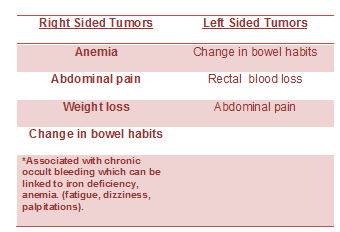
Colorectal cancer can present as asymptomatic and symptomatic. Asymptomatic patients are diagnosed with a fecal occult blood test are identified at an early stage and most commonly located in the cecum/ascending colon.
Symptoms differ depending on where the tumor is located.
A diagnosis is made by a Colorectal Cancer screening examination or through an evaluation for an unrelated illness.
Associated Co-morbidities[edit | edit source]
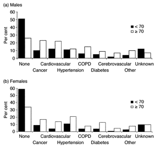
Approximately 51-59% of individuals with a diagnosis of CRC who are under the age of 70 do not also suffer from co-morbidities; however, in the individuals who are greater than 70 years of age, only 26-24% of them do not suffer from co-morbidities. Of this group greater than 70 years of age, the men have the highest prevalence of complicating co-morbid conditions. These conditions can have a marked impact on the treatment of the individual’s CRC diagnosis. The short-term survival is also worsened in the presence of co-morbid conditions, especially cardiovascular co-morbidities.
- Cardiovascular disease
- Previously diagnosed CA
- Male – Large Bowel, Urinary Tract, Lung, and Prostate
- Female – Large Bowel, Breast, and Female Genital System
- Hypertension (F>M)
- Chronic Obstructive Pulmonary Disease
- Diabetes
Medications[edit | edit source]
Chemotherapy
Targeted Drug Therapy
Monoclonal Antibody Therapy: proteins engineered to help the body’s natural immune system to attack and destroy colorectal cancer cells. It can be used independently or with other chemotherapy treatment.
Radiation Therapy: helps to destroy cancer cells and can be used in conjunction with chemotherapy.
Radiation options:
Intensity Modulated Radiation Therapy (IMRT)
Intraoperative Radiation Therapy (IORT)
CyberKnife®
TheraShpere®
TomoTherapy®
Trilogy™
Diagnostic Tests/Lab Tests/Lab Values[1][edit | edit source]
Preventative Scopes
- Flexible Sigmoidoscopy Exam: This test looks at the inner lining of the large intestine and is used for patients with abdominal pain, rectal bleeding, changes in bone, and people who are greater than 50 years of age. This is less invasive than a colonoscopy and does not require anesthesia.
- Colonoscopy: This test is used to check for polyps and the paitent’s risk for developing CRC. It is performed by the patient ingesting a laxative that results in bowel elimination and then the patient is put under an anesthesia. A scope is then inserted into the colon for visualizing the colon. Removal and biopsies can also be done during this procedure.
Blood Tests
- Complete Blood Count (CBC): This is used to check for anemia or too few red blood cells. This can occur in CRC because of prolonged bleeding from the tumor.
- Liver Enzymes: This is used to check the function of the live, due to the liver being a common organ for CRC to metastasize.
- Tumor Markers: CRC cells can sometimes produce bi-products that are released into the bloodstream. Two examples of these are Carcinoembryonic Antigen (CEA and CA 19-9). Commonly the tumor marker blood tests are used in conjunction with other tests to monitor individual’s treatment progress as well as an early sign that a cancer has returned. This is not used to screen or diagnose CRC, because not all CRC’s will show a release of tumor markers and some results may show up abnormal but are due to other disease processes, such as ulcerative colitis, non-cancerous tumors of intestines, or types of liver disease or chronic lung disease, or smoking.
Biopsy
- A biopsy is normally done if any other diagnostic test has suspected CRC. A small piece of tissue is removed through a scope during a colonoscopy. The tissue sample is tested in a lab by a pathologist under a microscope. This is the only way to determine for certain that the suspected tissue is in fact colorectal cancer.
- Biopsied tissue can also be tested for specific gene changes in the cancer cells that have an effect on the way the cancer is treated, for example, the KRAS and BRAF genes. Those two genes specifically have a large impact on the type of cancer treatment those patients receive and respond to.
- Biopsied tissue may be tested for changes called microsatellite instability (MSI). This is commonly present in hereditary non-polyposis colon cancer (HNPCC), as well as, some cancers not caused by HNPCC. If this is found in a cancer patient, because it is hereditary, family members may want to be tested also.
Computerized Tomography Scan (CT or CAT)
- This imaging test is an x-ray that produces detailed cross-sectional images of the body. This machine takes many pictures of the body as they are moving and creates detailed images of the soft tissues of the body. This test is usually done to help determine if the cancer has spread to the live or other organs.
- CT with portography: This is done to specifically look at the portal vein or the vein that goes from the liver to the intestines to look for the spread of the cancer to the liver.
- CT- guided needle biopsy: This is done when a suspected area of cancer lies deep within the body and a biopsy is taken using the imaging for location of the needle. This too often shows tumors in the liver.
Ultrasound
- This imaging test uses sound waves and their echo to create a picture of internal organs or masses. These can be done to look for tumors in the liver, gallbladder, pancreas, or anywhere else in the abdomen. This test cannot be done to look for tumors of the colon.
- Endorectal ultrasound: Specialized ultrasound used to evaluate colon and rectal cancers. The ultrasound transducer is inserted directly into the rectum. This is used to detect how far through the rectal wall the cancer has penetrated and if it has spread to nearby organs or lymph nodes.
- Intraoperative ultrasound: This specialized ultrasound is done during surgery while the abdomen is open. The transducer is placed directly on the liver and used to detect the spread of colorectal cancer to the liver.
Magnetic Resonance Imaging (MRI) scan
- This imaging scan provides detailed images of the soft tissues in the body. This imaging test uses radio waves and strong magnets instead of x-rays like the CT scan. A contrast material called gadolinium can also be used to see more precise images. This test is used to look at areas of the liver, where rectal cancer may have spread and also nearby structures to the colon and rectum. Endorectal MRI can also be used to improve the accuracy of this imaging test.
Chest X-ray
- This test may be ordered by a MD to detect if CRC has spread to the lung tissue.
Positron Emission Tomography (PET) scan
- This imaging test is done by injecting a form of a radioactive sugar (low radioactivity) into the blood. This sugar is absorbed by cancer cells because they grow rapidly in the body. A picture is then made of your body, highlighting the areas of radioactivity in your body. This test provides helpful information about your entire body. This test may be ordered to see if abnormal areas are tumors, to see if cancer has spread to the lymph nodes or if the MD feels the cancer has spread, but does not know for sure where it has spread.
Angiography
- This is an imaging test that uses an x-ray procedure to look at blood vessels. This is done by injecting a contrast dye into an artery before the x-ray picture is taken. This may be done to look at the arteries that supply blood to tumors in the liver as well as helping to plan the surgical removal of a tumor in the liver.
Etiology/Causes[edit | edit source]
Colon cancer originates from rapid cell proliferation of the epithelial cells called colonocytes that line the bowel, and somatic mutations in the p53 tumor-suppressor gene. The majority of CRCs are believed to occur sporadically leaving only about 10% to 20% of CRCs to have a known hereditary component
Developing polyps is a potential risk factor for CRC, particularly if they are adenomatous (glandular hyperplasia). The age of when polyps are diagnosed can be an indicator for prognosis and risk for developing cancer. Patients with adenomatous colorectal polyps have an increased risk of 1.78 in developing CRC. The risk increases if the polyps are diagnosed before the age of 60 to 2.59. The larger the polyp the greater probability the polyp is cancerous compared to smaller polyps. Another kind of adenomatous polyp is called familial adenomatous polyposis which is an autosomal-dominant disease. This disease has an occurrence rate of 1:7000 to 1:10,000. The colon is completely covered with polyps and if medical management does not take action approximately 50-75% of patient will develop CRC.
Risks
Genetic influence of relatives who have been diagnosed with any type of cancer can increase the risk for CRC. A first-degree relative with CRC has a 2-4 times the risk. Cancer family syndrome is an autosomal-dominant dis
order that puts the patient at 33% risk of developing cancer by the age of 50.
Genetic changes are responsible and have an important role for hyperproliferation of carcinogenic cells which due to ulcerative colitis, acromegaly, family history of colonic neoplasia, certain professions, smoking & drinking, consumption of red or processed meat, etc.
Inflammation of the colon and rectum are associated with an increased risk of colorectal cancer. The large intestines have an increase amount of bacteria present compared to the small intestines. This is can become problematic as it relates to an increase in proliferation of colonic carcinogens. Bile acids secreted in the intestines can act as tumor promoters and has shown to contribute to colon cancer.
Twenty-five percent of patients who have inflammatory bowel disease, ulcerative colitis, have an increased risk if they’ve had the disease for over 25 years. At a lesser degree, Crohn’s disease may also have an influence of risk.
Systemic Involvement[edit | edit source]
add text here
Medical Management (current best evidence)[edit | edit source]
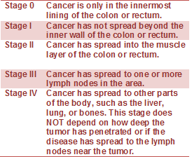
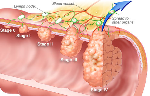
Staging of Colorectal Cancer
If cancer is detected, it will be "staged," a process of finding out how far the cancer has spread. Tumor size may not correlate with the stage of cancer. Staging also enables your doctor to determine what type of treatment you will receive.
Physical Therapy Management (current best evidence)[edit | edit source]
add text here
Alternative/Holistic Management (current best evidence)[edit | edit source]
Nutrition Therapy
Preventing Colorectal Cancer: Diet
There are steps you can take to dramatically reduce your odds of developing colorectal cancer. Researchers estimate that eating a nutritious diet, getting enough exercise, and controlling body fat could prevent 45% of colorectal cancers. The National Cancer Institute recommends a low-fat diet that includes plenty of fiber and at least five servings of fruits and vegetables per day.
-solid food may not be an option, depending on the stage and treatment
Pain management
Naturopathic therapy
Mind Body Medicine
Differential Diagnosis[edit | edit source]
add text here
Case Reports/ Case Studies[edit | edit source]
add links to case studies here (case studies should be added on new pages using the case study template)
Resources
[edit | edit source]
Recent Related Research (from Pubmed)[edit | edit source]
see tutorial on Adding PubMed Feed
Failed to load RSS feed from http://eutils.ncbi.nlm.nih.gov/entrez/eutils/erss.cgi?rss_guid=1NGmwZeh8JwVIzrKgHG1LrDm0izTr7ViJiDkSYAY2BW5hiXsx0|charset=UTF-8|short|max=10: Error parsing XML for RSS
References[edit | edit source]
- ↑ 1.0 1.1 Colorectal Cancer Overview [Internet]. American Cancer Society. 2013 [updated 2013 Jan 17]. Available from: http://www.cancer.org/cancer/colonandrectumcancer/overviewguide/colorectal-cancer-overview-key-statistics.
- ↑ 2.0 2.1 Seer Stat Facts Sheets: Colon and Rectum [Internet]. National Cancer Institute. 2012 [updated 2011 Nov]. Available from: http://seer.cancer.gov/statfacts/html/colorect.html.
- ↑ Enzinger PC, Benson AB, Mitchell EP, et al. Medical Update on Colorectal Cancer Understanding KRAS [pamphlet]. New York: Elsevier Oncology; 2010.
- ↑ Colorectal (Colon) Cancer [Internet]. Center for Disease Control. 2012 [updated 2012 Oct 22]. Available from: http://www.cdc.gov/cancer/colorectal/index.htm.
- ↑ Tidy, Colin MD. Colorectal Cancer [Internet]. Patient.co.uk; 2012 [updated 2012 July 19]. Available from: http://www.patient.co.uk/doctor/colorectal-adenocarcinoma.htm.
- ↑ De Marco MF, Janssen-Heijnen ML, Van Der Heijden LH, et al. Comorbidity and colorectal cancer according to subsite and stage: a population-based study. Eur J Cancer. 2000 Jan; 36(1): 95-9
