Chest X-Rays
Original Editor - Sweta Christian Top Contributors - Sweta Christian, Eki Tunjungsari, Rachael Lowe, Kim Jackson, Adam Vallely Farrell, Karen Wilson, Samuel Winter and Admin
Purpose
[edit | edit source]
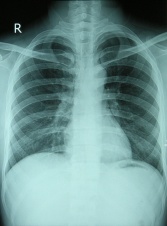
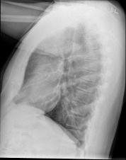
Chest X-rays is a painless, non-invasive test and is the most commonly preferred diagnostic examination to produce images of heart, lungs, airways, blood vessels and the bones of the spine and chest[1][2].
Technique
[edit | edit source]
An X-ray uses electromagnetic waves and ionizing radiation to create pictures of the inside of your body.
The procedures involves positioning the body between the machine that produces the X-rays and a plate that that creates the image digitally or with X-ray film[1].
- Front view:
- Posterior - Anterior (PA). This is the most common and preferred ype of chest X-Ray. Posterior - anterior refers to the direction of the X-Ray beam travel.; i e. X-Ray beams hit the posterior part of the chest before the anterior part. To obtain the image, the patient is asked to stand with their chest against the film, to hold their arms up or to the sides and roll their shoulders forward. The X-ray technician may then ask the patient to take few deep breaths and hold it for a couple of seconds. This techniques of holding the breath generally helps to get a clear picture of the heart and lungs on the image.
- Anterior - posterior (AP): This type of chest X-Ray is generally less preferred because the image of the heart and mediastinum is less clear and focused in this projection. To obtain AP image, the patient is asked to stand with their back against the film. If the patient is unable to stand, an AP image can also be taken with the patient sitting or supine on the bed.
- Decubitus: This type of X-Ray shows a frontal view of the chest when the patient is lying on their side(s). Decubitus X-Ray can be performed to assess the air inside the lung, the presence of free fluid, or airway obstruction.
2, For the side-view: The patient is asked to turn and place one shoulder on the plate and raise their hands over their head. The technician may again ask the patient to take a deep breath and hold it.
Analysis[edit | edit source]
Basic Checks[edit | edit source]
Reading the chest X-ray systematically reduces the chance of missed diagnosis. There is no one recommended analysis methodology; some may choose to read a chest X-Ray in an anatomical order and some may choose to use a mnemonic. However, everyone should begin analysing an X-Ray by ensuring the following details are correct:
- Patient's full name and date of birth
- Date and time of the X-Ray taken. There could be a few images taken on the same day.
- Image Annotation
- Right (R) or Left (left side)
- Image projection: PA (posterior-anterior) or AP (anterior-posterior) or lateral
- Patient's position. Check whether the patient is upright, semi-erect, or supine when the image was taken.
- Image Quality (R.I.P)
- R - Rotation. Check whether the patient's position is rotated. The patient is NOT rotated if the spinous processes are equidistant from the medial ends of the clavicles.
- I - Inspiration. Check whether the 5 - 7 anterior ribs intersect the diaphragm in the mid-clavicular line. If the intersection happens in anterior ribs 8 or more, then the lungs are hyperinflated.
- P - Penetration. Check whether the spine can be seen through the heart; which indicates that the X-ray exposure is adequate.
- Is there any previous films for comparison purposes?
- Foreign objects such as lines, tubes, pacemaker, drains, etc. Foreign objects can also be non-medical such as bullets, glass, etc.
Systematic Assessment[edit | edit source]
The most commonly used mnemonic approach to assess Chest X-Ray is the following:
| Mnemonic | Explanation | Note |
|---|---|---|
| A | Airways | The trachea should be central or slightly to the right. If it is deviated, check whether it is due to the patient's position or another pathological cause. |
| B | Bones and soft tissues | |
| C | Cardiac | |
| D | Diaphragm | |
| E | (Pleural) Effusion | |
| F | Fields and Fissure | |
| G | Great Vessels | |
| H | Hilum and mediastinum | |
| I |
The following video explains how to analyse a Chest- x-ray in nutshell
Evidence[edit | edit source]
A study conducted by Tape and Mushlin (1988) to study the effect of routine chest x-rays of pre-operative patients at risk for postoperative disease. Patient records from 341 admissions were reviewed to determine the relationship between chest x-ray results and postoperative chest complications. Patients who had major abnormalities had a 40% postoperative complication rate, compared with 9% for those with normal x-rays; but only 13% of the complications occurred in patients with major abnormalities. Nine patients had x-ray findings that led to clinical action: three with potentially beneficial management changes (congestive heart failure in 2, fibrosis in 1) and six with potentially detrimental clinical action (false diagnosis of tuberculosis in 2, false diagnosis of nodules in 2, falsely normal chest x-ray in 2). None of 50 surgical cancellations occurred as a result of an abnormal x-ray. All the beneficial effects attributable to preoperative chest x-rays accrued to patients who had clinical evidence of chest disease. The authors conclude that routine chest x-rays were not helpful in improving patient outcomes. They recommend ordering preoperative chest x-rays based on clinical indications so that the likelihood of false positives and false negatives and their associated detrimental effects can be minimized[3].
Resources
[edit | edit source]
How to read a Chest X-ray?
http://www.med.upenn.edu/normalcxr/
References[edit | edit source]
- ↑ 1.0 1.1 RadiologyInfo.org. X-Ray (Radiography) - Chest.http://www.radiologyinfo.org/en/info.cfm?pg=chestrad
- ↑ National Heart, Lung, and Blood Institute. Chest X-Ray. https://www.nhlbi.nih.gov/health/health-topics/topics/cxray
- ↑ Tape TG, Mushlin AI. How useful are routine chest x-rays of preoperative patients at risk for postoperative chest disease?. Journal of general internal medicine 1988; 3.1: 15-20.


