Cervical Myelopathy
Original Editor - Bridgit A Finley.
Lead Editors - Your name will be added here if you are a lead editor on this page. Read more.
Clinically Relevant Anatomy
[edit | edit source]
Chronic cervical degeneration is the most common cause of progressive spinal cord and nerve root compression. Spondylotic changes can result in stenosis of the spinal canal, lateral recess, and foramina. Spinal canal stenosis can lead to myelopathy, whereas the latter two can lead to radiculopathy.
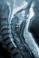
Osteophytes with cord compression
Mechanism of Injury / Pathological Process
[edit | edit source]
The onset is incidious and gradual and is related to degenerative changes in the cervical spine anatomy. Osteophytic overgrowth, thickning of the ligamentum flavum (dorsally) and of the posterior longitudional ligament can compress the spinal cord. The intervertebral discs dry out resulting in loss of disc height, which increase compression of the vertebrae/end plates and osteophytic spurs develop at the margins of the end plates. The degenerative changes encroach on the spinal cord and cause compression.
Clinical Presentation[edit | edit source]
Cervical spondylotic myelopathy can cause a variety of signs and symptoms. The onset is insidious, which typically becomes apparent in persons aged 50-60 years. About half of patients with cervical myelopathy have pain in their neck, scapular area or arms; most have symptoms of arm and leg dysfunction. Arm symptoms may include weakness, numbness (nonspecific/dermathomal) or clumsiness in the hands. Leg symptoms may include weakness, difficulty walking, and/or frequent falls. In later cases, bladder and bowel incontinence can occur. The first signs are often increased knee and ankle reflexes. A Myelopathy is an upper motor neuron lesion and the patients may present with spasticity, hyperreflexia, clonus, Babinski and Hoffman's sign.
Sex Both sexes are affected equally. Cervical spondylosis usually starts earlier in men (50 years) than in women (60 years).
Age 40-60 years old. Radiologic spondylotic changes increase with patient age; 70% of asymptomatic persons older than 70 years have some form of degenerative change in the cervical spine.
Diagnostic Procedures[edit | edit source]
MRI of the cervical spine can identify spinal canal stenosis, as well as rule out spinal cord tumors. Plane radiographs alone are of little use as an initial diagnostic procedure.
Neck Disability Index[edit | edit source]
http://academic.regis.edu/clinicaleducation/pdf's/NDI_with_scoring.pdf
Management / Interventions
[edit | edit source]
add text here relating to management approaches to the condition
Differential Diagnosis
[edit | edit source]
Special Tests: (+) Clonus, (+) Hoffman's Sign
(+) Lhermitte's Sign - flexion of the cervical spine may cause an "electric shock" sensation down the center of the back.
Common Symptoms
Distal weakness, decreased ROM in the cervical spine, especially extension.
Clumsy or weak hands
Pain in shoulder or arms
Unsteady gait
Increased reflexes in the lower extremities and in the upper extremities below the level of the lesion.
MRI may be useful to diagnose myelopathy. Electromyography (EMG) and nerve conduction velocity (NCV) may help rule out peripheral nerve radiculopathy.
Key Evidence[edit | edit source]
add text here relating to key evidence with regards to any of the above headings
Resources
[edit | edit source]
add appropriate resources here
Case Studies[edit | edit source]
add links to case studies here (case studies should be added on new pages using the case study template)
Recent Related Research (from Pubmed)[edit | edit source]
Failed to load RSS feed from http://eutils.ncbi.nlm.nih.gov/entrez/eutils/erss.cgi?rss_guid=10SuWQ8ymABjulX7yzOMA4mM0Ok422wmPAwrVWPUMJAoZUZ9gK|charset=UTF-8|short|max=10: Error parsing XML for RSS
References[edit | edit source]
References will automatically be added here, see adding references tutorial.


