Cerebral Cortex: Difference between revisions
No edit summary |
No edit summary |
||
| Line 8: | Line 8: | ||
The cerebral cortex | The cerebral cortex | ||
* Represents in humans a highly developed structure concerned with the most familiar functions we associate with the human brain. Between 14 billion and 16 billion neurons are found in the cerebral cortex.<ref name=":1" /> | * Represents in humans a highly developed structure concerned with the most familiar functions we associate with the human brain. Between 14 billion and 16 billion neurons are found in the cerebral cortex.<ref name=":1" /> | ||
* Highly convoluted external surface of the brain. Its distinctive shape arose during evolution as the volume of the cortex increased more rapidly than the cranial volume resulting in the convolution of the surface and the folding of the total structure of the cortex. | * Highly convoluted external surface of the brain. Its distinctive shape arose during evolution as the volume of the cortex increased more rapidly than the cranial volume resulting in the convolution of the surface and the folding of the total structure of the cortex. If the cerebral cortex were to be removed and unfolded, it would cover several yards or meters. | ||
* The convolutions consist of grooves known as sulci that separate the more elevated regions called gyri. | * The convolutions consist of grooves known as sulci that separate the more elevated regions called gyri. | ||
* The cortex has been divided into four lobes using certain consistently present sulci as landmarks. These lobes are named after the overlying cranial bones: frontal, parietal, temporal and occipital<ref>[http://www.ifc.unam.mx/Brain/cercox.htm Nervous system] Available from:http://www.ifc.unam.mx/Brain/cercox.htm (accessed 25.12.2020)</ref>. | * The cortex has been divided into four lobes using certain consistently present sulci as landmarks. These lobes are named after the overlying cranial bones: frontal, parietal, temporal and occipital<ref>[http://www.ifc.unam.mx/Brain/cercox.htm Nervous system] Available from:http://www.ifc.unam.mx/Brain/cercox.htm (accessed 25.12.2020)</ref>. | ||
== Structure == | |||
[[File:The brain 1.png|right|frameless]] | |||
The outer layer of the cerebral hemisphere is termed the cerebral cortex. This is inter-connected via pathways that run sub-cortically. It is these connections as well as the connections from the cerebral cortex to the brainstem, spinal cord and nuclei deep within the cerebral hemisphere that form the white matter of the cerebral hemisphere. The deep nuclei include structures such as the basal ganglia and the thalamus. | |||
The cerebrum consists of two cerebral hemispheres, the right and left hemisphere are connected by the corpus callosum which facilitates communication between both sides of the brain, with each hemisphere in the main connection to the contralateral side of the body i.e. the left hemisphere of the cerebrum receives information from the right side of the body resulting in motor control of the right side of the body and vice versa. | |||
The hemispheres are divided into four lobes; | |||
# Occipital | |||
# Parietal | |||
# Temporal (medial part of which are a series of structures including the Hippocampus) | |||
# Frontal | |||
== Neocortex == | == Neocortex == | ||
[[File:Neocortex.png|right|frameless]] | The neocortex is the largest and most powerful area of the human brain. All of its important cognitive functions are made possible by the convergence of two distinct streams of information: a "bottom-up" stream, which represents signals from the environment, and a "top-down" stream, which transmits internally generated information about past experiences and current aims.<ref>medicalxpress [https://medicalxpress.com/news/2020-11-region-brain-key-source-encoding.html Researchers identify a region of the brain as a key source of signals encoding past experiences in the neocortex] Available from: https://medicalxpress.com/news/2020-11-region-brain-key-source-encoding.html (accessed 25.12.20200</ref>[[File:Neocortex.png|right|frameless]] | ||
The phylogenetically most recent part of the cerebral cortex, the neocortex, has six horizontal layers (the more ancient part of the cerebral cortex, the hippocampus, has at most three cellular layers). Neurons in various layers connect vertically to form small microcircuits, called 'columns'. | The phylogenetically most recent part of the cerebral cortex, the neocortex, has six horizontal layers (the more ancient part of the cerebral cortex, the hippocampus, has at most three cellular layers). Neurons in various layers connect vertically to form small microcircuits, called 'columns'. | ||
| Line 54: | Line 66: | ||
# Sensory areas: receive input from the thalamus and process information related to the senses. They include the visual cortex of the occipital lobe, the auditory cortex of the temporal lobe, the gustatory cortex, and the somatosensory cortex of the parietal lobe. Within the sensory areas are association areas that give meaning to sensations and associate sensations with specific stimuli. | # Sensory areas: receive input from the thalamus and process information related to the senses. They include the visual cortex of the occipital lobe, the auditory cortex of the temporal lobe, the gustatory cortex, and the somatosensory cortex of the parietal lobe. Within the sensory areas are association areas that give meaning to sensations and associate sensations with specific stimuli. | ||
2. Motor areas: including the primary motor cortex and the premotor cortex, regulate voluntary movement<ref name=":1" />. | 2. Motor areas: including the primary motor cortex and the premotor cortex, regulate voluntary movement<ref name=":1" />. | ||
[[File:Wrinkles.jpg|right|frameless]] | |||
== | == Why Wrinkles are Good! == | ||
Over time, the human cortex undergoes a process of corticalization, or wrinkling of the cortex. This process is due to the vast knowledge that the human brain accumulates over time. Therefore, the more wrinkly your brain, the smarter and more intelligent you are!<ref>Brain made simple [https://brainmadesimple.com/cerebral-cortex-and-lobes-of-the-brain/ Cerebral Cortex] Available from:https://brainmadesimple.com/cerebral-cortex-and-lobes-of-the-brain/ (accessed 25.12.2020)</ref> | |||
or | |||
== References == | == References == | ||
Revision as of 08:06, 25 December 2020
Original Editor - Lucinda hampton
Top Contributors - Lucinda hampton, Rucha Gadgil and Joao Costa
Introduction[edit | edit source]
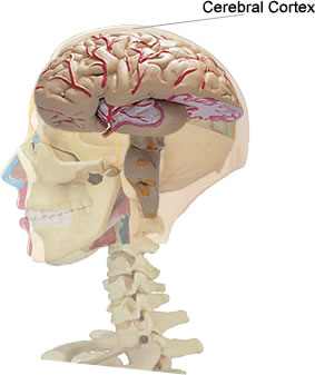
The cerebral cortex is a sheet of neural tissue that is outermost to the cerebrum of the mammalian brain. It has up to six layers of nerve cells. It is covered by the meninges and often referred to as gray matter[1]. The cortex is gray because nerves in this area lack the insulation (myelin) that makes most other parts of the brain appear to be white[2].
The cerebral cortex
- Represents in humans a highly developed structure concerned with the most familiar functions we associate with the human brain. Between 14 billion and 16 billion neurons are found in the cerebral cortex.[2]
- Highly convoluted external surface of the brain. Its distinctive shape arose during evolution as the volume of the cortex increased more rapidly than the cranial volume resulting in the convolution of the surface and the folding of the total structure of the cortex. If the cerebral cortex were to be removed and unfolded, it would cover several yards or meters.
- The convolutions consist of grooves known as sulci that separate the more elevated regions called gyri.
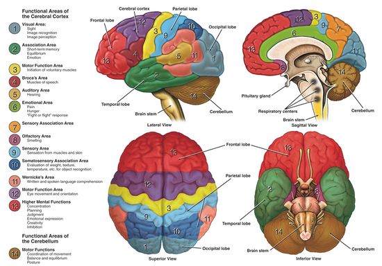
- The cortex has been divided into four lobes using certain consistently present sulci as landmarks. These lobes are named after the overlying cranial bones: frontal, parietal, temporal and occipital[3].
Structure[edit | edit source]
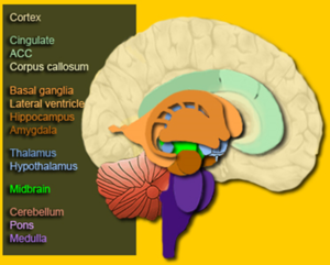
The outer layer of the cerebral hemisphere is termed the cerebral cortex. This is inter-connected via pathways that run sub-cortically. It is these connections as well as the connections from the cerebral cortex to the brainstem, spinal cord and nuclei deep within the cerebral hemisphere that form the white matter of the cerebral hemisphere. The deep nuclei include structures such as the basal ganglia and the thalamus.
The cerebrum consists of two cerebral hemispheres, the right and left hemisphere are connected by the corpus callosum which facilitates communication between both sides of the brain, with each hemisphere in the main connection to the contralateral side of the body i.e. the left hemisphere of the cerebrum receives information from the right side of the body resulting in motor control of the right side of the body and vice versa.
The hemispheres are divided into four lobes;
- Occipital
- Parietal
- Temporal (medial part of which are a series of structures including the Hippocampus)
- Frontal
Neocortex[edit | edit source]
The neocortex is the largest and most powerful area of the human brain. All of its important cognitive functions are made possible by the convergence of two distinct streams of information: a "bottom-up" stream, which represents signals from the environment, and a "top-down" stream, which transmits internally generated information about past experiences and current aims.[4]
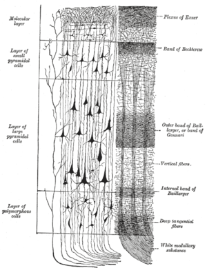
The phylogenetically most recent part of the cerebral cortex, the neocortex, has six horizontal layers (the more ancient part of the cerebral cortex, the hippocampus, has at most three cellular layers). Neurons in various layers connect vertically to form small microcircuits, called 'columns'.
Image:Cerebral cortex. To the left, the groups of cells; to the right, the systems of fibers. Quite to the left of the figure a sensory nerve fiber is shown. Cell body layers are labeled on the left, and fiber layers are labeled on the right.
- The neocortex is the newest part of the cerebral cortex to evolve. The six-layer neocortex is a distinguishing feature of mammals; it has been found in the brains of all mammals, but not in any other animals. In humans, 90% of the cerebral cortex is neocortex.
- In humans, 90% of the cerebral cortex and 76% of the entire brain is neocortex[5][1]
Allocortex[edit | edit source]
The allocortex (also known as heterogenetic cortex) is a part of the cerebral cortex characterized by fewer cell layers than the neocortex (i.e. fewer than six). More ancient phylogenetically than the mammals, evolved to handle olfaction and the memory of smells.
The specific regions of the brain normally described as part of the allocortex are:
Allocortex: fewer than six layers, more ancient phylogenetically than the mammals, evolved to handle olfaction and the memory of smells.
- Archicortex
- Olfactory cortex
- Hippocampus
2. Paleocortex (3 three to five layers)
The cellular organization of the old cortex is different from the six-layer structure mentioned above. Unable to form so many complex micro circuits.
Function[edit | edit source]
The cerebral cortex is involved in several functions of the body including:
Determining intelligence
Determining personality
Motor function
Planning and organization
Touch sensation
Processing sensory information
Language processing
The cerebral cortex contains:
- Sensory areas: receive input from the thalamus and process information related to the senses. They include the visual cortex of the occipital lobe, the auditory cortex of the temporal lobe, the gustatory cortex, and the somatosensory cortex of the parietal lobe. Within the sensory areas are association areas that give meaning to sensations and associate sensations with specific stimuli.
2. Motor areas: including the primary motor cortex and the premotor cortex, regulate voluntary movement[2].
Why Wrinkles are Good![edit | edit source]
Over time, the human cortex undergoes a process of corticalization, or wrinkling of the cortex. This process is due to the vast knowledge that the human brain accumulates over time. Therefore, the more wrinkly your brain, the smarter and more intelligent you are![6]
References[edit | edit source]
- ↑ 1.0 1.1 Kidzsearch Cerebral Cortex Available from:https://wiki.kidzsearch.com/wiki/Cerebral_cortex (accessed 25.12.2020)
- ↑ 2.0 2.1 2.2 Thought Co. What Does the Brain's Cerebral Cortex Do? Available from:https://www.thoughtco.com/anatomy-of-the-brain-cerebral-cortex-373217 (accessed 25.12.2020)
- ↑ Nervous system Available from:http://www.ifc.unam.mx/Brain/cercox.htm (accessed 25.12.2020)
- ↑ medicalxpress Researchers identify a region of the brain as a key source of signals encoding past experiences in the neocortex Available from: https://medicalxpress.com/news/2020-11-region-brain-key-source-encoding.html (accessed 25.12.20200
- ↑ Science Daily Neocortex Available from:https://www.sciencedaily.com/terms/neocortex.htm (accessed 26.12.2020)
- ↑ Brain made simple Cerebral Cortex Available from:https://brainmadesimple.com/cerebral-cortex-and-lobes-of-the-brain/ (accessed 25.12.2020)


