Basal Ganglia
This article or area is currently under construction and may only be partially complete. Please come back soon to see the finished work! (12/01/2020)
Original Editor - Your name will be added here if you created the original content for this page.
Top Contributors - Lucinda hampton, Lauren Heydenrych, Kim Jackson, Tony Lowe, Joao Costa, Rucha Gadgil and Aminat Abolade
Introduction[edit | edit source]
The “basal ganglia” refers to a group of subcortical nuclei responsible primarily for motor control, as well as other roles such as motor learning, executive functions and behaviors, and emotions. Proposed more than two decades ago, the classical basal ganglia model shows how information flows through the basal ganglia back to the cortex through two pathways with opposing effects for the proper execution of movement[1].
Much of the model has remains today however the model has been modified and amplified with the emergence of new data. The basal ganglia network is now viewed as multiple parallel loops and re-entering circuits whereby motor, associative, and limbic territories are engaged mainly in the control of movement, behavior, and emotions. These parallel circuits have been found that subserve the other functions of the basal ganglia engaging associative and limbic territories.[1]
Disruption of the basal ganglia network forms the basis for several movement disorders.
Structure[edit | edit source]
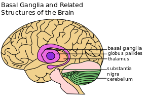
The basal ganglia are a cluster of subcortical nuclei deep to cerebral hemispheres. The largest component of the basal ganglia is the corpus striatum which contains the caudate and lenticular nuclei (the putamen, globus pallidus externus, and internus), the subthalamic nucleus (STN), and the substantia nigra (SN). These structures intricately synapse onto one another to promote or antagonize movement.[2]
Corpus Striatum- (The largest subcortical brain structure of the basal ganglia is the striatum with a volume of approximately 10 cm). Comprises divided into a ventral striatum, and a dorsal striatum, subdivisions that are based upon function and connections. The ventral striatum consists of the nucleus accumbens and the olfactory tubercle. The dorsal striatum consists of the caudate nucleus and the putamen. A white matter, nerve tract (the internal capsule) in the dorsal striatum separates the caudate nucleus and the putamen.[4] Anatomically, the term striatum describes its striped (striated) appearance of grey-and-white matter.[2]
Subthalamic Nucleus (STN) -
Substantia Nigra (SN)
Sub Heading 3[edit | edit source]
Resources[edit | edit source]
- bulleted list
- x
or
- numbered list
- x
References[edit | edit source]
- ↑ 1.0 1.1 Lanciego JL, Luquin N, Obeso JA. Functional neuroanatomy of the basal ganglia. Cold Spring Harbor perspectives in medicine. 2012 Dec 1;2(12):a009621.Available from:https://www.ncbi.nlm.nih.gov/pmc/articles/PMC3543080/ (last accessed 12.1.2020)
- ↑ 2.0 2.1 Young CB, Sonne J. Neuroanatomy, basal ganglia. InStatPearls [Internet] 2018 Dec 28. StatPearls Publishing.Available from:https://www.ncbi.nlm.nih.gov/books/NBK537141/ (last accessed 12.1.2020)


