Aponeurosis
Original Editor - User Name
Top Contributors - Lucinda hampton, Kim Jackson and Ahmed M Diab
Introduction[edit | edit source]
Aponeuroses are sheet-like elastic tendon structures that cover a portion of the muscle belly and act as insertion sites for muscle fibers while free tendons connect muscles to bones[1]. They have a role similar to a tendon but here is how they differ:
- An aponeurosis looks quite different than a tendon. An aponeurosis is made of layers of delicate, thin sheaths. Tendons, in contrast, are tough and rope-like. An aponeurosis is made primarily of bundles of collagen fibers distributed in regular parallel patterns, which makes an aponeurosis resilient.
- Aponeurosis has a function of absorbing energy during the movement of the muscle, while Tendon has a function of stretching and contracting during muscle movements.
- It is very rare for the Aponeurosis to get injured as it is situated hidden under many layers of bones and muscles. But Tendon gets injured easily, for it is present in all the injury-prone areas.
- Aponeuroses can act as fascia. Fascia is a fibrous tissue that envelopes muscles or organs, to bind muscles together or to other tissues.[2][3].
Sub Heading 2[edit | edit source]
The erector spinae aponeurosis is a common aponeurosis that blends with the thoracolumbar fascia, with a proximal attachment on the sacrum an the spinous processes of the lumbar vertebrae, for the three erector spinae muscles (iliocostalis, longissimus, and spinalis) and overlying the inferior portion of the erector spinae muscles.[4]
Sub Heading 3[edit | edit source]
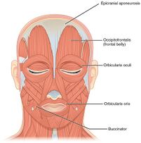
The epicranial (or galea) aponeurosis is a tough fibrous sheet of connective tissue that extends over the cranium, forming the middle (third) layer of the scalp. The epicranial aponeurosis also contains vessels that communicate between the deep vascular plexus contained within the subgaleal layer below as well as the superficial vascular plexus in the subcutaneous layer above[5].
Image : epicranial aponeurosis.
Resources[edit | edit source]
- bulleted list
- x
or
- numbered list
- x
References[edit | edit source]
- ↑ Arellano CJ, Gidmark NJ, Konow N, Azizi E, Roberts TJ. Determinants of aponeurosis shape change during muscle contraction. Journal of biomechanics. 2016 Jun 14;49(9):1812-7. Available:https://pubmed.ncbi.nlm.nih.gov/27155748/ (accessed 15.12.2021)
- ↑ Study.com Aponeurosis Available: https://study.com/academy/lesson/aponeurosis-definition-function.html (accessed 15.12.2021)
- ↑ Ask any difference Aponeurosis and tendon Available:https://askanydifference.com/difference-between-aponeurosis-and-tendon/ (accessed 15.12.2021)
- ↑ IMAIOS Erector spinae aponeurosis - Aponeurosis musculis erectoris spinae Available:https://www.imaios.com/en/e-Anatomy/Anatomical-Parts/erector-spinae-aponeurosis (accessed 15.12.2021)
- ↑ Radiopedia galea Aponeurosis Available:https://radiopaedia.org/articles/galea-aponeurotica (accessed 15.12.2021)


