Ankle and Foot Fractures: Difference between revisions
No edit summary |
No edit summary |
||
| Line 1: | Line 1: | ||
== Search Strategy == | == Search Strategy == | ||
We searched scientific articles and information by using PubMed, web of knowledge, Google Scholar, PEDro and Medline. We used keywords such as ankle and foot fractures, Lis franc fracture, malleolar fracture, calcaneus fracture, surgical or operative treatment and nonsurgical or conservative treatment, ankle sprain and ankle fracture. We also used some inclusion criteria as foot and ankle fractures and exclusion criteria as ankle sprain or ankle instability to find relevant articles in French or English. | We searched scientific articles and information by using PubMed, web of knowledge, Google Scholar, PEDro and Medline. We used keywords such as ankle and foot fractures, Lis franc fracture, malleolar fracture, calcaneus fracture, surgical or operative treatment and nonsurgical or conservative treatment, ankle sprain and ankle fracture. We also used some inclusion criteria as foot and ankle fractures and exclusion criteria as ankle sprain or ankle instability to find relevant articles in French or English. | ||
== Definition/Description == | == Definition/Description == | ||
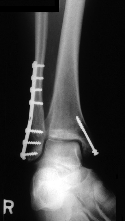
An ankle fracture is a fracture in a bone that shapes the ankle. This can be the superior articular surface of the talus, the end of the fibula (malleolus lateralis), the end of the tibia (malleolus medialis) or both (bimalleolar fracture) (Figure 1). They usually result from an external or internal rotation injury to the ankle. <br> Ankle fractures can range from the less serious avulsion injuries (small pieces of bone that are pulled off) to severe shattering-type breaks. The ankle ligaments, keeping the ankle in its normal position, could possibly be involved. These fractures are among the most common fractures in adults. (7) Ankle fractures are common musculoskeletal injuries that occur in a bimodal distribution. These fractures are common in younger men and older women, the former related to high-energy trauma and the latter to osteopenia and osteoporosis. (29) | |||
Ankle fractures are usually caused by falls, twisting injuries and sports injuries. (4)<br> | |||
<br> | |||
== Clinically Relevant Anatomy == | == Clinically Relevant Anatomy == | ||
Revision as of 12:29, 30 May 2016
Search Strategy[edit | edit source]
We searched scientific articles and information by using PubMed, web of knowledge, Google Scholar, PEDro and Medline. We used keywords such as ankle and foot fractures, Lis franc fracture, malleolar fracture, calcaneus fracture, surgical or operative treatment and nonsurgical or conservative treatment, ankle sprain and ankle fracture. We also used some inclusion criteria as foot and ankle fractures and exclusion criteria as ankle sprain or ankle instability to find relevant articles in French or English.
Definition/Description[edit | edit source]
An ankle fracture is a fracture in a bone that shapes the ankle. This can be the superior articular surface of the talus, the end of the fibula (malleolus lateralis), the end of the tibia (malleolus medialis) or both (bimalleolar fracture) (Figure 1). They usually result from an external or internal rotation injury to the ankle.
Ankle fractures can range from the less serious avulsion injuries (small pieces of bone that are pulled off) to severe shattering-type breaks. The ankle ligaments, keeping the ankle in its normal position, could possibly be involved. These fractures are among the most common fractures in adults. (7) Ankle fractures are common musculoskeletal injuries that occur in a bimodal distribution. These fractures are common in younger men and older women, the former related to high-energy trauma and the latter to osteopenia and osteoporosis. (29)
Ankle fractures are usually caused by falls, twisting injuries and sports injuries. (4)
Clinically Relevant Anatomy[edit | edit source]
add text here
Epidemiology /Etiology[edit | edit source]
There are different causes for an ankle fracture:
- A big force that works into the bone. For example a kick or a smack during sport activities or a car accident.
- A little piece of the bone tears off when there is pulled at a ligament. For example when you stumble.
- Twisting or rotating your ankle
- Rolled your ankle
Characteristics/Clinical Presentation[edit | edit source]
- Difficulties or even inability to walk or load the ankle. (it is possible to walk with less severe breaks, so never rely on walking as a test of whether a bone has been fractured.
- Pain
- Swelling, along the length of the leg or more localized
- Blisters (over the fracture site).
- Bruising (soon after the injury).
- Difference in appearance.
When an ankle has been broken, there is not only structural damage to the skeletal structure, but also to the ligament tissue (deltoid ligament and the anterior and posterior tibiofibular ligaments) and possibly nervous and musculoskeletal tissue around the ankle complex.. This can result in impaired balance capacity, reduced joint position sense, slowed nerve conduction, velocity, impaired cutaneous sensation and decreased dorsal extension range of motion[1]
Differential Diagnosis[edit | edit source]
add text here
Diagnostic Procedures[edit | edit source]
To evaluate the ankle in the acute fase, we use the ‘Ottawa ankle rules’. Ottawa_Ankle_Rules
Examination[edit | edit source]
add text here related to physical examination and assessment
Medical Management
[edit | edit source]
Most patients with a malleolus fracture require 6 weeks of immobilization. Patients with an initially non-displaced fracture or who were treated surgically will generally require 4 weeks of non-weight bearing in a short-leg cast or removable walking boot, followed by 2 weeks in a walking cast or boot. The removable boot will allow for earlier range-of-motion exercises.
Surgery is needed for many types of ankle fractures. While not always necessary, surgery for ankle fractures is not uncommon. The need for surgery depends on the appearance of the ankle joint on X-ray and the type of ankle fracture.
Adequate reduction with congruency of the joint has been reported as one of the most important indications of a good end result. Inadequate reduction may lead to osteoarthritis.
Physical Therapy Management
[edit | edit source]
After 6 weeks of immobilization, the ankle can be fully loaded. There is no standardized rehabilitation program after cast removal. Each program is individually[1]
Physiotherapists are often involved in the rehabilitation, which starts quickly (1 week) after the period of immobilization. Most people experience pain, swelling, stiffness, muscle atrophy and decreased muscle torque[2], impaired ankle mobility, impaired balance capacity and increased ankle circumference[1] at the ankle after cast removal. Consequently, patients complain of limitation in activities involving the lower limb, such as stair climbing, walking and reduced participation in work and recreation. It has been found that patients with unimalleolar fractures report less activity limitation than those with bimalleolar or trimalleolar fractures[3]
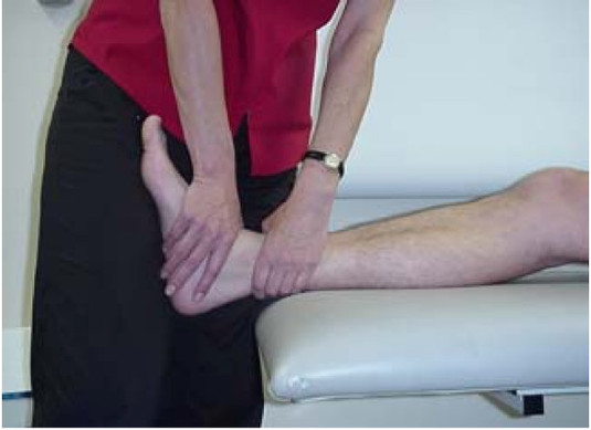
Passive joint mobilization is commonly used to work on the problems of pain and joint stiffness, in order to allow an earlier return to activities. For this technique, the physiotherapist manually glides the articular surfaces of a joint to produce oscillatory movements. It has been proven that manual therapy, such as joint mobilization, produces analgesic effects. It also increases elasticity of joint structures through interactions at the local, central nervous system and psychological levels[4]
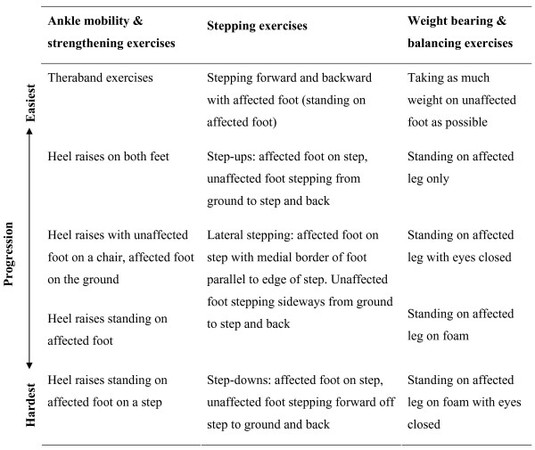
There is evidence that, after a surgical treatment for an ankle fracture, a training program, started within one week after cast removal and continued for 12 weeks (with 2 appointments per week), shows superior results compared to usual care, regarding patient scored function and muscle strength in the plantar flexors and dorsiflexors of patients under the age of 40. The patients had to do home exercises daily, prescribed by the physiotherapist, appropriate to the functional status at the time. Functional goals are loaded ankle dorsiflexion, plantairflexion, on-leg-stance, rising on toes, rising on heels, normalized walking pattern when walking on even ground, on stairs and at comfortable speed[5]
When the cast is removed, many patients have a plantarflexion contracture (http://www.physio-pedia.com/index.php5?title=Plantarflexion_contracture). This contracture is not caused directly by fracture but develops as an adaptive response to immobilization. The addition of a program of passive stretches has no benefit over exercise alone for the treatment of plantarflexion contracture after cast immobilization[6]
Resources
[edit | edit source]
http://orthopedics.about.com/od/footanklefractures/Information_About_Foot_Ankle_Fractures.htm
http://www.foothealthfacts.org/footankleinfo/ankle-fracture.htm
http://www.invaliditeit.be/enkelbreuk.html
http://www.associatie-orthopedie-lier.be/Generic/servlet/Main.html?p_pageid=37245
http://www.dokterdokter.nl/medisch/folder/view/id/1582/gebroken-enkel
Clinical Bottom Line[edit | edit source]
add text here
Recent Related Research (from Pubmed)[edit | edit source]
see tutorial on Adding PubMed Feed
Extension:RSS -- Error: Not a valid URL: Feed goes here!!|charset=UTF-8|short|max=10
References [edit | edit source]
- ↑ 1.0 1.1 1.2 Nilsson G, Nyberg P, Ekdahl CH, Eneroth M. Performance after surgical treatment of patients with ankle fractures – 14-month follow-up. Physiotherapy Research International 2003, 8(2) 69-82
- ↑ Nilsson G, Jonsson K, Ekdahl CH, Eneroth M. Outcome and quality of life after surgically treated ankle fractures in patients 65 years or older. BMC Musculoskeletal Disorders 2007;8:127
- ↑ Lin CC, Moseley AM, Herbert RD, Refshauge KM. Pain and dorsiflexion range of motion predict short-and medium-term activity limitation in people receiving physiotherapy intervention after ankle fracture: an observational study. Australian Journal of Physiotherapy 2009, 55;31-37
- ↑ Lin C CH, Moseley AM, Refshauge KM, Haas M, Herbert RD. Effectiveness of joint mobilisation after cast immobilisation for ankle fracture: a protocol for a randomised controlled trial. BMC Musculoskeletal Disorders 2006;7:46
- ↑ Nilsson G, Jonsson K, Ekdahl CH, Eneroth M. Effects of a training program after surgically treated ankle fracture : a prospective randomised controlled trial. BMC Musculoskeletal Disorders 2009;10:118
- ↑ Moseley AM, Herbert RD, Nightingale EJ, Taylor DA, Evans TM, Robertson GJ, Gupta SK, Penn J. Passive stretching does not enhance outcomes in patients with plantarflexion contracture after cast immobilization for ankle fracture : a randomized controlled trial. Arch Phys Med Rehabil 2005;86 :1118-26


