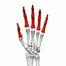
Phalangeal Fractures
Introduction[edit | edit source]
Fractures of the finger bones, known as phalanges, frequently occur and are often seen in emergency departments and clinics[1]. These injuries can affect the proximal, middle, or distal phalanx. In most cases of phalanx fractures, effective realignment can be achieved through non-surgical methods. Timely intervention is crucial to promote healing and restore functionality.
Clinical Anatomy[edit | edit source]
The hand's proximal and middle phalanges share a common anatomical structure comprising a head, neck, shaft, and base. Meanwhile, the distal phalanx is characterized by its tuft, shaft, and base divisions. The proximal phalanx is stabilized by surrounding structures, including proper and accessory collateral ligaments, the volar plate, and extensor/flexor tendons. The middle phalanx has two primary insertions: the central slip (part of the extensor mechanism) and the flexor digitorum superficialis (FDS). In the anatomy of the distal phalanx, the distal interphalangeal joint (DIPJ) is surrounded by extensor and flexor tendons, the volar plate, and collateral ligaments. The flexor digitorum profundus (FDP) inserts at the volar metaphysis of the distal phalanx. At the proximal interphalangeal joint (PIPJ), the flexor digitorum profundus and the flexor digitorum superficialis share a sheath. The flexor digitorum superficialis lies on the volar side, while the flexor digitorum profundus is on the dorsal side. As these tendons traverse the PIPJ, the flexor digitorum superficialis bifurcates into two slips, forming the Camper's chiasm, which inserts on the volar aspect of the middle phalanx. This significant anatomical relationship can result in a swan neck deformity, characterized by a hyperextended PIPJ and a flexed DIPJ.
Etiology[edit | edit source]
Phalangeal fractures of the hand are usually the result of a direct trauma, crush or twisting injury[2]
Clinical Presentation[edit | edit source]
Patients with metacarpal fractures generally present with[3]
- Tenderness
- swelling
- bruising
- crepitus
- deformity
- restricted motion and instability are common signs of injury
References[edit | edit source]
- ↑ Clarence Kee; Patrick Massey. Phalanx fracture. InStatPearls [Internet]2023 August 14. StatPearls Publishing.Available from:https://www.ncbi.nlm.nih.gov/books/NBK545182/
- ↑ Laura Kremer,Johannes Frank,Thomas Lustenberger,Ingo Marzi &Anna Lena Sander, Epidemiology and treatment of phalangeal fractures: conservative treatment is the predominant therapeutic concept, European Journal of Trauma and Emergency Surgery, Springer, Published online: 25 May 2020, volume 48, pages 567-571 (2022)
- ↑ Benjamin J.F Dean, Christopher Little.Fractures of the metacarpals and phalanges.Orthopaedic and Trauma volume 25,Issue 1, February 2011,Pages 43-56


