File:Anterior view of posterolateral attachments of lateral menicus.jpeg

Size of this preview: 673 × 599 pixels. Other resolution: 1,890 × 1,683 pixels.
Original file (1,890 × 1,683 pixels, file size: 608 KB, MIME type: image/jpeg)
Summary[edit | edit source]
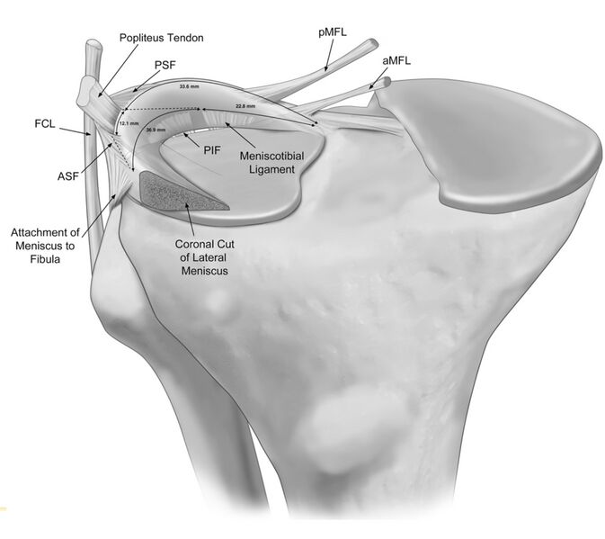
Anterior view illustration of the anatomic attach- ments to the posterolateral meniscus. The meniscotibial liga- ment and the posteroinferior popliteomeniscal fascicle (PIF) are depicted emerging from posterior to the lateral tibial pla- teau and attaching to the inferior margin of the posterolateral meniscus. aMFL, anterior meniscofemoral ligament; pMFL, posterior meniscofemoral ligament; ASF, anterosuperior popliteomeniscal fascicle; PSF, posterosuperior popliteome- niscal fascicle; FCL, fibular collateral ligament.
Licensing[edit | edit source]
The copyright holder has given special permission to use this work in Physiopedia.
File history
Click on a date/time to view the file as it appeared at that time.
| Date/Time | Thumbnail | Dimensions | User | Comment | |
|---|---|---|---|---|---|
| current | 17:03, 31 March 2023 | | 1,890 × 1,683 (608 KB) | Robert Pierce (talk | contribs) | Anterior view illustration of the anatomic attach- ments to the posterolateral meniscus. The meniscotibial liga- ment and the posteroinferior popliteomeniscal fascicle (PIF) are depicted emerging from posterior to the lateral tibial pla- teau and attaching to the inferior margin of the posterolateral meniscus. aMFL, anterior meniscofemoral ligament; pMFL, posterior meniscofemoral ligament; ASF, anterosuperior popliteomeniscal fascicle; PSF, posterosuperior popliteome- niscal fascicle; FCL, fi... |
You cannot overwrite this file.
File usage
The following page uses this file:

