Glioblastoma
Original Editors - Simone Potts from Bellarmine University's Pathophysiology of Complex Patient Problems project.
Lead Editors - Your name will be added here if you are a lead editor on this page. Read more.
Definition/Description[edit | edit source]
An astrocytoma is a glioma that develops from star-shaped glial cells (astrocytes) that support nerve cells. A glioblastoma multiforme is classified as a grade IV astrocytoma. It is also referred to as a glioblastoma or GBM[1]
Glioblastoma multiforme (GBM) is by far the most common and most malignant of the glial tumors. Attention was recently drawn to this form of brain cancer when Senator Ted Kennedy was diagnosed with glioblastoma and ultimately died from it.
Gliomas comprise a heterogeneous group of neoplasms that differ in location within the central nervous system, in age and sex distribution, in growth potential, in extent of invasiveness, in morphological features, in tendency for progression, and in response to treatments.
GBM can spread through the brain tissue, but rarely spreads to other areas outside of the central nervous system.
All GBM tumors have abnormal and numerous blood vessels, a common feature of a fast-growing tumor. These blood vessels deliver necessary oxygen and nutrients to GBM tumors, helping them grow and spread. In addition, these blood vessels easily mix with normal brain tissue and travel away from the main tumor, which makes GBM tumors a challenge to treat.[2]
Prevalence[edit | edit source]
add text here
Characteristics/Clinical Presentation[edit | edit source]
add text here
Associated Co-morbidities[edit | edit source]
add text here
Medications[edit | edit source]
add text here
Diagnostic Tests/Lab Tests/Lab Values[edit | edit source]
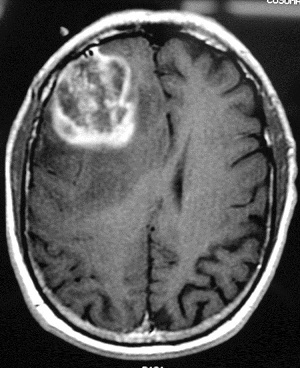
T1-weighted axial gadolinium-enhanced magnetic resonance image demonstrates an enhancing tumor of the right frontal lobe. Image courtesy of George Jallo, MD.[3]
Etiology/Causes[edit | edit source]
add text here
Systemic Involvement[edit | edit source]
add text here
Medical Management (current best evidence)[edit | edit source]
add text here
Physical Therapy Management (current best evidence)[edit | edit source]
add text here
Alternative/Holistic Management (current best evidence)[edit | edit source]
add text here
Differential Diagnosis[edit | edit source]
add text here
Case Reports/ Case Studies[edit | edit source]
add links to case studies here (case studies should be added on new pages using the case study template)
Resources
[edit | edit source]
add appropriate resources here
Recent Related Research (from Pubmed)[edit | edit source]
see tutorial on Adding PubMed Feed
Extension:RSS -- Error: Not a valid URL: Feed goes here!!|charset=UTF-8|short|max=10


