Synovial Joints
Original Editor - Lucinda hampton
Top Contributors - Lucinda hampton and Joao Costa
Introduction[edit | edit source]
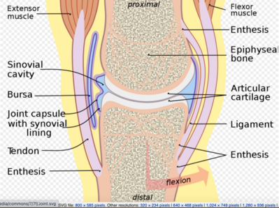
Synovial joints are the most common type of joint in the body (see image 1.). A key structural characteristic for a synovial joint that is not seen at fibrous or cartilaginous joints is the presence of a joint cavity. The joint cavity contains synovial fluid, secreted by the synovial membrane (synovium), which lines the articular capsule. This fluid-filled space is the site at which the articulating surfaces of the bones contact each other. Hyaline cartilage forms the articular cartilage, covering the entire articulating surface of each bone. The articular cartilage and the synovial membrane are continuous. A few synovial joints of the body have a fibrocartilage structure located between the articulating bones. This is called an articular disc, which is generally small and oval-shaped, or a meniscus, which is larger and C-shaped.[1][2].
Synovial joints are often further classified by the type of movements they permit. There are six such classifications: hinge (elbow), saddle (carpometacarpal joint), planar (acromioclavicular joint), pivot (atlantoaxial joint), condyloid (metacarpophalangeal joint), and ball and socket (hip joint).[1]
Sub Heading 2[edit | edit source]
Sub Heading 3[edit | edit source]
Resources[edit | edit source]
- bulleted list
- x
or
- numbered list
- x
References[edit | edit source]
- ↑ 1.0 1.1 Juneja P, Hubbard JB. Anatomy, Joints. 2018 Available: https://www.ncbi.nlm.nih.gov/books/NBK507893/ (accessed 20.6.2021)
- ↑ OSE Synovial joints Available:https://open.oregonstate.education/aandp/chapter/9-4-synovial-joints/ (accessed 20.6.2021)


