Strength-Duration Curve
Introduction[edit | edit source]
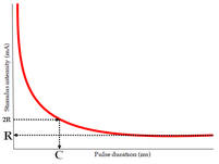
The strength-duration curve (SDC) is a graphical representation of the relationship between the intensity of an electrical stimulus at the motor point of a muscle and the length of time taken to elicit a minimal contraction in that muscle.
Clinical Significance[edit | edit source]
It ascertains the excitability of the nerve and thus, can detect the magnitude of nerve damage. It can show recovery over a period of time. It is a valuable diagnostic and prognostic tool. It is usually performed after 3 weeks of nerve injury to allow for Wallerian degeneration.
Types of Strength-Duration Curve[edit | edit source]
Normal Innervation[edit | edit source]
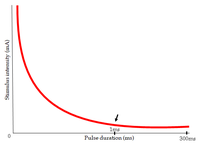
This is also called as a "nerve curve". All nerve fibers supplying the muscle are intact. The shape of the curve is a continuous rectangular hyperbola. The same intensity is required to produce a response at longer durations. The intensity increases steadily for shorter durations. The curve is usually seen rising at the 1ms mark.
Complete Denervation[edit | edit source]
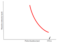
This is also called as a "muscle curve". All nerve fibers supplying the muscle are degenerated. Th curve is characteristically steep and shifted to the right. The intensity keeps increasing when lowering the duration below 100ms. There is no response seen at very short durations.
Partial Denervation[edit | edit source]
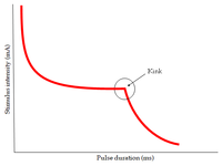
Some of the nerve fibers supplying the muscles are degenerated while others are intact. A characteristic kink is present in the curve. Right side of the curve represents the denervated part of the muscle while the left side represents the innervated fibers of the muscle. Higher intensity stimulates denervated muscle fibers and lower intensity stimulates innervated muscle fibers.
Rheobase[edit | edit source]
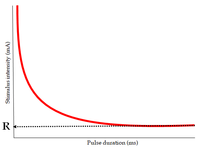
It is the minimum intensity of current required to stimulate a muscle at infinite duration. Its normal value ranges between 2 and 18 mA. The rheobase is greater for denervated muscles.
Chronaxie[edit | edit source]
It is the minimum time required for a current of double the intensity of rheobase to stimulate a muscle. Its normal value is below 1ms. Chronaxie is inversely proportional to excitability. Thus, its value is greater for denervated muscles.
Utilization time[edit | edit source]
It is the time taken by a stimulus of rheobasic strength to excite the nerve and produce a muscle contraction. Below this value, there will be no muscle contraction.
Factors affecting the Strength-Duration Curve[edit | edit source]
- Skin resistance
- Subcutaneous tissue like fat
- Temperature
- Electrode size, material and placement
- Age of the subject
- Fatigue
Advantages of the Strength-Duration Curve[edit | edit source]
It is quick and easy to perform. It requires minimal training. It is economical in comparison to other clinical tests.
Disadvantages of the Strength-Duration Curve[edit | edit source]
It provides only qualitative data in relation to the degree of denervation. It cannot locate the site of the lesion. In large muscles, only few fibers can be studied due to the limits of the method.
Reference[edit | edit source]
Forster A, Palastanga N, editors. Clayton's Electrotherapy. 8th edition. New Delhi: CBS Publishers. 2005
