Medial Patellofemoral Ligament (MPFL)
This article or area is currently under construction and may only be partially complete. Please come back soon to see the finished work! (17/11/2020)
Original Editor - Beverly Klinger
Top Contributors - Beverly Klinger
Description[edit | edit source]
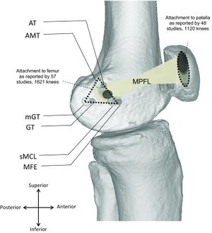
The Medial Patellofemoral Ligament (MPFL) is an hour-glass shaped ligament made of bands of retinacular tissue. The MPFL plays a significant role in the stabilization of the medial aspect of the patella. Especially during the early stages of knee flexion, the MPFL is a critical component in patellar tracking and stability within the trochlear groove .
Attachments[edit | edit source]
The medial patellofemoral ligament is located in the second layer of three soft tissue layers within the medial aspect of the knee. The MPFL originates from a triangular space running between the adductor tubercle, medial femoral condyle and gastconemius tubercle, superior to the superficial medial collateral ligament (MCL)[1][2] The MPFL inserts onto the superomedial aspect of the patella[1]. The proximal insertion extends to the quadriceps tendon while the distal insertion crosses deep to the distal vastus medialis obliquus (VMO)[2].
Function[edit | edit source]
The main function of the medial patellofemoral ligament is to provide restraint to the patella during early knee flexion (0-30 degrees)[2]. It acts in maintaining appropriate patellar tracking within the trochlear groove, while providing 50-60% of the restraining force against lateral displacement[1][2].
Clinical relevance[edit | edit source]
During acute lateral patellar dislocations (LPD), the MPFL is ruptured >90% of the time with almost 100% rupture occurring in repeat dislocations. Lateral dislocations most often occur when the foot is planted and an internal rotational force is applied to a flexed, valgus knee joint[2]. MPFL reconstruction surgery is often performed in patients with patellofemoral instability who have suffered recurrent lateral patellar dislocations. Surgical repair of the MPFL restores the medial patellar stability.[3]
Assessment[edit | edit source]
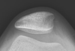
Medial patellofemoral ligament rupture will present with pain and tenderness along the medial retinaculum as well as the medial border of the patella. The patient will present with apprehension during lateral translation of the patella with the absence of a firm end feel. Radiographic images (lateral and sunrise view) will show bony contusions as well as lateral subluxation of the patella which would indicate a possible MPFL tear. [2] Magnetic Resonance Imaging (MRI) provides the most accurate assessment of MPFL soft tissue integrity.
Treatment[edit | edit source]
Conservative treatment, especially after the first lateral patellar dislocation, has been regarded as the most appropriate course of treatment. Rehabilitation with Physical Therapy and bracing is the prescribed treatment with surgical intervention of the MPFL when conservative treatment fails or the patient presents with recurring dislocations.[2]
References[edit | edit source]
- ↑ 1.0 1.1 1.2 Cite error: Invalid
<ref>tag; no text was provided for refs named:0 - ↑ 2.0 2.1 2.2 2.3 2.4 2.5 2.6 Cite error: Invalid
<ref>tag; no text was provided for refs named:1 - ↑ Cite error: Invalid
<ref>tag; no text was provided for refs named:2 - ↑ Nabil Ebraheim. Medial Patellofemoral Ligament Of The Knee Anatomy ,Everything You Need To Know - Dr. Nabil Ebraheim. Available from: https://www.youtube.com/watch?v=VFbe-PhnfJE. [Last accessed 19/11/20]


