File:Tinea Versicolor Microscope.JPG
No higher resolution available.
Tinea_Versicolor_Microscope.JPG (432 × 535 pixels, file size: 38 KB, MIME type: image/jpeg)
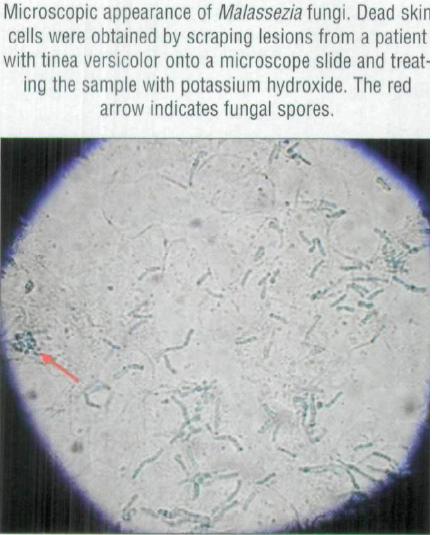
"Microscopic appearance of Malassezia fungi. Dead skin cells were obtained by scraping lesions from a patient with tinea versicolor onto a microscope siide and treating the sample with potassium hydroxide. The red arrow indicates fungal spores."
File history
Click on a date/time to view the file as it appeared at that time.
| Date/Time | Thumbnail | Dimensions | User | Comment | |
|---|---|---|---|---|---|
| current | 04:40, 11 April 2010 | | 432 × 535 (38 KB) | Jamie Rife (talk | contribs) | "Microscopic appearance of Malassezia fungi. Dead skin cells were obtained by scraping lesions from a patient with tinea versicolor onto a microscope siide and treating the sample with potassium hydroxide. The red arrow indicates fungal spores." |
You cannot overwrite this file.
File usage
The following page uses this file:

