Search results
Page title matches
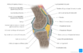
File:Knee joint - Kenhub.png Knee joint(1,400 × 896 (728 KB)) - 14:10, 16 March 2022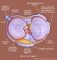
File:Knee-joint-meniscus.jpg (324 × 345 (29 KB)) - 21:17, 30 September 2013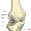
File:Knee joint anterior aspect Primal.png (660 × 660 (279 KB)) - 21:36, 7 December 2020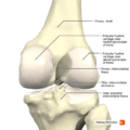
File:Knee joint posterior aspect Primal.png (660 × 660 (289 KB)) - 21:37, 7 December 2020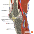
File:Sagittal section of the knee joint Primal.png (660 × 660 (607 KB)) - 21:46, 7 December 2020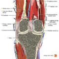
File:Coronal section of the knee joint 2 Primal.png (660 × 660 (629 KB)) - 21:45, 7 December 2020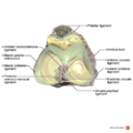
File:Ligaments of the knee joint superior aspect Primal.png (990 × 990 (409 KB)) - 21:37, 7 December 2020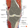
File:Coronal section of the knee joint 1 Primal.png (660 × 660 (658 KB)) - 21:44, 7 December 2020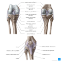
File:Overview of the knee joint (anterior and posterior views) - Kenhub.png Overview of the knee joint (anterior and posterior views)(1,400 × 1,400 (1.04 MB)) - 04:53, 30 March 2022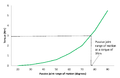
File:Torque displacement curve for the ankle joint with the knee extended.png (1,159 × 753 (16 KB)) - 01:15, 23 June 2019
File:Figure 3. Torque and displacement curve for the ankle joint with the knee extended.pdf Figure 3. Torque and displacement curve for the ankle joint with the knee extended(0 × 0 (94 KB)) - 00:46, 21 June 2019


_-_Kenhub.png)

Page text matches
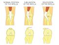
File:Patella Dislocation and Subluxation - Shutterstock Image - ID 637362388.jpg ...a or kneecap. Anatomy of human knee joint, front view of straight and bent knee. Aksanaku. Shutterstock.com(1,300 × 1,000 (382 KB)) - 12:06, 14 June 2022
File:Baker's Cyst - Shutterstock - ID 2163640049.jpg ...e, and joint with Popliteal cyst. The flow of synovial fluid from the knee joint to the gastrocnemio-semimembranosus bursa. vector illustration. Vector Form(2,231 × 1,997 (674 KB)) - 01:01, 24 May 2024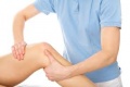
File:Figure 5.0.jpg Manual therapy at knee joint(276 × 183 (5 KB)) - 00:47, 8 May 2018
File:Heleen fig1.jpg anatomical structure of the knee joint, fig 1(800 × 484 (28 KB)) - 16:48, 30 December 2010
File:Condyles of femur LD.B.0190.004.L.jpg Condyles of femur. Interior of right knee joint, anterior view. Image #190-4(640 × 458 (110 KB)) - 18:48, 13 February 2020
File:Guideline for the management of knee and hip osteoarthritis .png management of hip and knee osteoarthritis. The objective of this guideline is to present the best ava other than joint replacement for the hip and knee.(1,920 × 1,080 (344 KB)) - 01:38, 26 February 2019File:Figure 3. Torque and displacement curve for the ankle joint with the knee extended.pdf Figure 3. Torque and displacement curve for the ankle joint with the knee extended(0 × 0 (94 KB)) - 00:46, 21 June 2019
File:Condyles of femur with cruciate ligaments LD.B.0190.005.L.jpg Interior of right knee joint, anterior cruciate ligament . Basset image ID Image #190-5(640 × 458 (111 KB)) - 18:51, 13 February 2020File:Knee joint - Kenhub.png Knee joint(1,400 × 896 (728 KB)) - 14:10, 16 March 2022File:Overview of the knee joint (anterior and posterior views) - Kenhub.png Overview of the knee joint (anterior and posterior views)(1,400 × 1,400 (1.04 MB)) - 04:53, 30 March 2022
File:Lateral HP prosthetic alignment.png ...al plane. The weight line or centre of gravity falls posterior to the knee joint and foot.(710 × 1,452 (420 KB)) - 14:48, 9 July 2022
File:Knee Joint.jpg Different views of the knee joint(2,250 × 1,725 (1.11 MB)) - 13:08, 24 February 2020File:Brunnstrom's Approach.pdf ...Synergy: Pelvis: anterior tilt Hip: Flexion, Abduction & External rotation Knee: Flexion Ankle: Dorsiflexion Toes: Extension ...t & fingers flexion Lower Limb Mixed Synergy: Pelvis post tilt hip add.+IR Knee extension Ankle & toes plantarflexion(0 × 0 (1.17 MB)) - 20:04, 2 January 2022


