File:Anterior view of posterolateral attachments of lateral menicus.jpeg: Difference between revisions
(Anterior view illustration of the anatomic attach- ments to the posterolateral meniscus. The meniscotibial liga- ment and the posteroinferior popliteomeniscal fascicle (PIF) are depicted emerging from posterior to the lateral tibial pla- teau and attaching to the inferior margin of the posterolateral meniscus. aMFL, anterior meniscofemoral ligament; pMFL, posterior meniscofemoral ligament; ASF, anterosuperior popliteomeniscal fascicle; PSF, posterosuperior popliteome- niscal fascicle; FCL, fi...) |
(added citation) |
||
| Line 1: | Line 1: | ||
== Summary == | == Summary == | ||
Anterior view illustration of the anatomic attach- ments to the posterolateral meniscus. The meniscotibial liga- ment and the posteroinferior popliteomeniscal fascicle (PIF) are depicted emerging from posterior to the lateral tibial pla- teau and attaching to the inferior margin of the posterolateral meniscus. aMFL, anterior meniscofemoral ligament; pMFL, posterior meniscofemoral ligament; ASF, anterosuperior popliteomeniscal fascicle; PSF, posterosuperior popliteome- niscal fascicle; FCL, fibular collateral ligament. | Anterior view illustration of the anatomic attach- ments to the posterolateral meniscus. The meniscotibial liga- ment and the posteroinferior popliteomeniscal fascicle (PIF) are depicted emerging from posterior to the lateral tibial pla- teau and attaching to the inferior margin of the posterolateral meniscus. aMFL, anterior meniscofemoral ligament; pMFL, posterior meniscofemoral ligament; ASF, anterosuperior popliteomeniscal fascicle; PSF, posterosuperior popliteome- niscal fascicle; FCL, fibular collateral ligament.<ref>Aman ZS, DePhillipo NN, Storaci HW, Moatshe G, Chahla J, Engebretsen L, et al. Quantitative and qualitative assessment of posterolateral meniscal anatomy: Defining the popliteal hiatus, popliteomeniscal fascicles, and the lateral meniscotibial ligament. Am J Sports Med [Internet]. 2019;47(8):1797–803. Available from: <nowiki>http://dx.doi.org/10.1177/0363546519849933</nowiki></ref> | ||
== Licensing == | == Licensing == | ||
{{allow-physiopedia}} | {{allow-physiopedia}} | ||
Revision as of 17:35, 31 March 2023
Summary[edit | edit source]
Anterior view illustration of the anatomic attach- ments to the posterolateral meniscus. The meniscotibial liga- ment and the posteroinferior popliteomeniscal fascicle (PIF) are depicted emerging from posterior to the lateral tibial pla- teau and attaching to the inferior margin of the posterolateral meniscus. aMFL, anterior meniscofemoral ligament; pMFL, posterior meniscofemoral ligament; ASF, anterosuperior popliteomeniscal fascicle; PSF, posterosuperior popliteome- niscal fascicle; FCL, fibular collateral ligament.[1]
Licensing[edit | edit source]
The copyright holder has given special permission to use this work in Physiopedia.
- ↑ Aman ZS, DePhillipo NN, Storaci HW, Moatshe G, Chahla J, Engebretsen L, et al. Quantitative and qualitative assessment of posterolateral meniscal anatomy: Defining the popliteal hiatus, popliteomeniscal fascicles, and the lateral meniscotibial ligament. Am J Sports Med [Internet]. 2019;47(8):1797–803. Available from: http://dx.doi.org/10.1177/0363546519849933
File history
Click on a date/time to view the file as it appeared at that time.
| Date/Time | Thumbnail | Dimensions | User | Comment | |
|---|---|---|---|---|---|
| current | 17:03, 31 March 2023 | 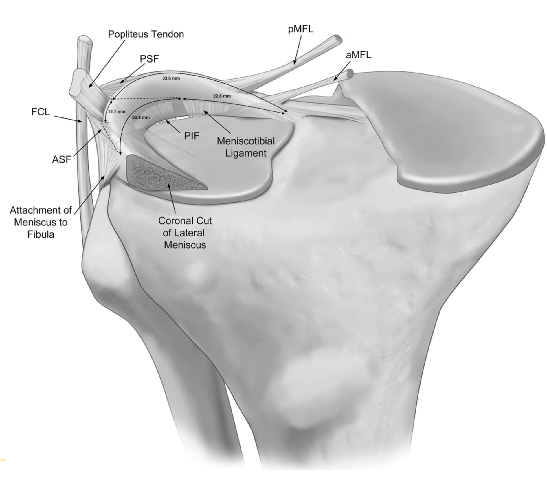 | 1,890 × 1,683 (608 KB) | Robert Pierce (talk | contribs) | Anterior view illustration of the anatomic attach- ments to the posterolateral meniscus. The meniscotibial liga- ment and the posteroinferior popliteomeniscal fascicle (PIF) are depicted emerging from posterior to the lateral tibial pla- teau and attaching to the inferior margin of the posterolateral meniscus. aMFL, anterior meniscofemoral ligament; pMFL, posterior meniscofemoral ligament; ASF, anterosuperior popliteomeniscal fascicle; PSF, posterosuperior popliteome- niscal fascicle; FCL, fi... |
You cannot overwrite this file.
File usage
The following page uses this file:

