Tracheobronchial Tree: Difference between revisions
mNo edit summary |
mNo edit summary |
||
| Line 14: | Line 14: | ||
The tracheobronchial tree has a branching structure of approximately 23 branches or generations extending from the trachea (generation 0) to the last order of terminal bronchioles (generation 23).<ref>Gauthier SP, Wolfson MR, Deoras KS, Shaffer TH. [https://www.nature.com/articles/pr199230.pdf?origin=ppub Structure-function of airway generations 0 to 4 in the preterm lamb.] Pediatric research. 1992 Feb;31(2):157-62.</ref><ref>Patwa A, Shah A. [https://www.ncbi.nlm.nih.gov/pmc/articles/PMC4613399/#:~:text=The%20terminal%20bronchioles%20(generation%2016,are%20completely%20lined%20with%20alveoli. Anatomy and physiology of respiratory system relevant to anaesthesia. Indian journal of anaesthesia]. 2015 Sep;59(9):533.</ref> | The tracheobronchial tree has a branching structure of approximately 23 branches or generations extending from the trachea (generation 0) to the last order of terminal bronchioles (generation 23).<ref>Gauthier SP, Wolfson MR, Deoras KS, Shaffer TH. [https://www.nature.com/articles/pr199230.pdf?origin=ppub Structure-function of airway generations 0 to 4 in the preterm lamb.] Pediatric research. 1992 Feb;31(2):157-62.</ref><ref>Patwa A, Shah A. [https://www.ncbi.nlm.nih.gov/pmc/articles/PMC4613399/#:~:text=The%20terminal%20bronchioles%20(generation%2016,are%20completely%20lined%20with%20alveoli. Anatomy and physiology of respiratory system relevant to anaesthesia. Indian journal of anaesthesia]. 2015 Sep;59(9):533.</ref> | ||
== Trachea == | == Trachea == | ||
[[File:Anatomy of trachea.png|thumb|400x400px|alt=|border| <small>''Figure 2: Anatomy of the trachea''</small> |left]]The main functions of the trachea | [[File:Anatomy of trachea.png|thumb|400x400px|alt=|border| <small>''Figure 2: Anatomy of the trachea''</small> |left]]The main functions of the trachea is to allow the passage of air into the lungs and provide a vital role in the mucociliary clearance of the respiratory system. | ||
Characteristics of the trachea | |||
# The trachea descends from the larynx into the lung.<ref name=":0" /> | # The trachea descends from the larynx into the lung.<ref name=":0" /> | ||
# The trachea is tube 12 cm.<ref>Brand-Saberi BE, Schäfer T. [https://pubmed.ncbi.nlm.nih.gov/24295654/ Trachea: anatomy and physiology. Thoracic surgery clinics]. 2014 Feb 1;24(1):1-5.</ref> | # The trachea is tube 12 cm.<ref>Brand-Saberi BE, Schäfer T. [https://pubmed.ncbi.nlm.nih.gov/24295654/ Trachea: anatomy and physiology. Thoracic surgery clinics]. 2014 Feb 1;24(1):1-5.</ref> | ||
# Is situated anterior to the oesophagus. | # Is situated anterior to the oesophagus. | ||
# .Extends from cricoid cartilage (c6 level) by cricothyroid membrane to carina (level of sternal angle). At the level of | # .Extends from cricoid cartilage (c6 level) by cricothyroid membrane to carina (level of sternal angle). | ||
# At the level of sternal angle, the trachea bifurcates into the right<nowiki/> and left main bronchi.<ref name=":0" /><ref name=":1" /> | |||
The trachea is a C-shaped structure, flexible tube that is composed of hyali<nowiki/>ne cartilage on the anterior and lateral walls. The posterior wall of the trachea is formed by the trachealis smooth muscle .Also the trachea is composed of several primary structural annular ligaments.<ref name=":1">Mieczkowski B, Seavey BF. [https://www.ncbi.nlm.nih.gov/books/NBK448070/ Anatomy, Head and Neck, Trachea.]</ref> (anatomy book ) | |||
'''Hyaline cartilage''' | |||
# Series of 16 to 20 hyaline cartilage rings | # Series of 16 to 20 hyaline cartilage rings connected by an annular ligament. | ||
# The tracheal cartilaginous rings, are responsible for keeping the trachea lumen open in spite of the changes in intrathoracic pressure that occur during respiration | # The tracheal cartilaginous rings, are responsible for keeping the trachea lumen open in spite of the changes in intrathoracic pressure that occur during respiration.<ref name=":1" /> | ||
===== '''Trachealis muscle''' ===== | ===== '''Trachealis muscle''' ===== | ||
| Line 34: | Line 39: | ||
* Is in contact with the anterior esophagus | * Is in contact with the anterior esophagus | ||
* The trachealis muscle functions to constrict the airway by pulling the cartilages together. | * The trachealis muscle functions to constrict the airway by pulling the cartilages together. By decreasing the diameter of the airway, it allows for increased expiratory force during coughing and thus allows greater velocity to be achieved for the particle to be expelled. This action is helpful, especially when eating food, which requires the expansion of the esophagus. | ||
* [[File:Cross section trachea.png|thumb|722x722px|alt=| ''<small>Figure 3: Cross-section of a trachea</small> <ref>https://commons.wikimedia.org/wiki/File:Cross_section_of_a_trachea_and_esophagus.svg </ref>''|center]] | * [[File:Cross section trachea.png|thumb|722x722px|alt=| ''<small>Figure 3: Cross-section of a trachea</small> <ref>https://commons.wikimedia.org/wiki/File:Cross_section_of_a_trachea_and_esophagus.svg </ref>''|center]] | ||
| Line 42: | Line 47: | ||
The trachea divides at the carina (at the level of the sternal angle) to give rise to the two primary bronchi – '''the right and left bronchus''' (figure X). Differences between right and left bronchus are illustrated below in table 1 .The right and left bronchus conduct air from the trachea into the right and left lung respectively. | The trachea divides at the carina (at the level of the sternal angle) to give rise to the two primary bronchi – '''the right and left bronchus''' (figure X). Differences between right and left bronchus are illustrated below in table 1 .The right and left bronchus conduct air from the trachea into the right and left lung respectively. | ||
The '''primary bronchi''' then branch into s'''econdary bronchi (lobar bronchi)''' – three on the right and two on the left. Each secondary bronchus supplies a lobe of | The '''primary bronchi''' then branch into s'''econdary bronchi (lobar bronchi)''' – three on the right and two on the left. Each secondary bronchus supplies a lobe of each lung. | ||
Each secondary bronchi (lobar bronchi) subsequently divide into narrower '''tertiary bronchi (segmental bronchi)''' that supply bronchopulmonary segments (largest subdivisions of a lobe). | Each secondary bronchi (lobar bronchi) subsequently divide into narrower '''tertiary bronchi (segmental bronchi)''' that supply bronchopulmonary segments (largest subdivisions of a lobe). | ||
| Line 56: | Line 61: | ||
- More vertical | - More vertical | ||
- Shorter (2 | - Shorter (2.5 cm) | ||
- Supported by C shaped cartilages | - Supported by C shaped cartilages | ||
| Line 80: | Line 85: | ||
'''Conducting bronchioles (part of conducting zone)''' | '''Conducting bronchioles (part of conducting zone)''' | ||
* | * Segmental bronchi give rise to 20 to 25 generations of conducting bronchioles that further divide to terminal bronchioles. | ||
* | * Function - as part of the conducting system they allow air to travel from the trachea to alveoli where gas exchange occurs. | ||
* | |||
* | '''Terminal bronchioles (part of conducting zone)''' | ||
* Represent the last part of the conducting part of the bronchial tree and give rise to several generations of respiratory bronchioles | |||
* | * Function - as part of the conducting system they allow air to travel from the trachea to alveoli where gas exchange occurs. | ||
* | |||
'''Respiratory bronchioles ( part of respiratory zone)''' | |||
* Distinguishable to other types of bronchioles by the presence of alveoli extending from their lumens | |||
* Function - represent the first part of the respiratory airways where they facilitate gas exchange | |||
Gas exchange occurs in the respiratory bronchioles, alveolar ducts and alveoli, which collectively form the respiratory zones deep in the lungs. | |||
Resources | |||
{{#ev:youtube|5F8XdE1d0xk|500}}<ref>Meditay.Trachea, Bronchial Tree and Alveolar Tree (Parts, Structures and Walls) - Anatomy Available from:https://www.youtube.com/watch?v=5F8XdE1d0xk</ref> | {{#ev:youtube|5F8XdE1d0xk|500}}<ref>Meditay.Trachea, Bronchial Tree and Alveolar Tree (Parts, Structures and Walls) - Anatomy Available from:https://www.youtube.com/watch?v=5F8XdE1d0xk</ref> | ||
== References == | == References == | ||
Revision as of 22:42, 22 April 2022
Original Editor - User Name
Top Contributors - Stella Constantinides and Kim Jackson
Introduction[edit | edit source]

The tracheobronchial tree is a branching tree of airways composed of:
- the trachea
- the bronchi
- the bronchioles.[2]
Its main function is the transfer of air from the environment to the lungs for gas exchange.
The tracheobronchial tree has a branching structure of approximately 23 branches or generations extending from the trachea (generation 0) to the last order of terminal bronchioles (generation 23).[3][4]
Trachea[edit | edit source]

The main functions of the trachea is to allow the passage of air into the lungs and provide a vital role in the mucociliary clearance of the respiratory system.
Characteristics of the trachea
- The trachea descends from the larynx into the lung.[2]
- The trachea is tube 12 cm.[5]
- Is situated anterior to the oesophagus.
- .Extends from cricoid cartilage (c6 level) by cricothyroid membrane to carina (level of sternal angle).
- At the level of sternal angle, the trachea bifurcates into the right and left main bronchi.[2][6]
The trachea is a C-shaped structure, flexible tube that is composed of hyaline cartilage on the anterior and lateral walls. The posterior wall of the trachea is formed by the trachealis smooth muscle .Also the trachea is composed of several primary structural annular ligaments.[6] (anatomy book )
Hyaline cartilage
- Series of 16 to 20 hyaline cartilage rings connected by an annular ligament.
- The tracheal cartilaginous rings, are responsible for keeping the trachea lumen open in spite of the changes in intrathoracic pressure that occur during respiration.[6]
Trachealis muscle[edit | edit source]
- Type of muscle : Smooth muscle
- Connects the ends of the C-shaped tracheal cartilages.
- Is in contact with the anterior esophagus
- The trachealis muscle functions to constrict the airway by pulling the cartilages together. By decreasing the diameter of the airway, it allows for increased expiratory force during coughing and thus allows greater velocity to be achieved for the particle to be expelled. This action is helpful, especially when eating food, which requires the expansion of the esophagus.
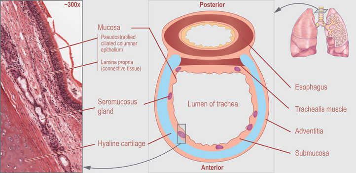 Figure 3: Cross-section of a trachea [7]

Bronchi[edit | edit source]
At the level of the fifth thoracic vertebra, the trachea divides into the left and right main bronchi. The right main bronchus has a length of 2.5 cm, and the left main bronchus is 5 cm long.
The trachea divides at the carina (at the level of the sternal angle) to give rise to the two primary bronchi – the right and left bronchus (figure X). Differences between right and left bronchus are illustrated below in table 1 .The right and left bronchus conduct air from the trachea into the right and left lung respectively.
The primary bronchi then branch into secondary bronchi (lobar bronchi) – three on the right and two on the left. Each secondary bronchus supplies a lobe of each lung.
Each secondary bronchi (lobar bronchi) subsequently divide into narrower tertiary bronchi (segmental bronchi) that supply bronchopulmonary segments (largest subdivisions of a lobe).
Segmental bronchi undergo further branching to form numerous smaller airways, called the bronchioles.
| Right Bronchus | Left Bronchus |
| - Wider
- More vertical - Shorter (2.5 cm) - Supported by C shaped cartilages - 20 to 30 degree angle |
- Narrower
- More angular - Longer (5cm) - Supported by C shaped cartilages - 40 to 60 degree angle |
Table 1
Bronchioles[edit | edit source]
Bronchioles arise from segmental bronchi and represent the smaller branches of the bronchial tree. They differ histologically from bronchi as they lack C shaped cartilages in their walls.
Bronchioles initially divide into many generations of conducting bronchioles which eventually divide further into terminal bronchioles which further divide into respiratory bronchioles.
Conducting bronchioles (part of conducting zone)
- Segmental bronchi give rise to 20 to 25 generations of conducting bronchioles that further divide to terminal bronchioles.
- Function - as part of the conducting system they allow air to travel from the trachea to alveoli where gas exchange occurs.
Terminal bronchioles (part of conducting zone)
- Represent the last part of the conducting part of the bronchial tree and give rise to several generations of respiratory bronchioles
- Function - as part of the conducting system they allow air to travel from the trachea to alveoli where gas exchange occurs.
Respiratory bronchioles ( part of respiratory zone)
- Distinguishable to other types of bronchioles by the presence of alveoli extending from their lumens
- Function - represent the first part of the respiratory airways where they facilitate gas exchange
Gas exchange occurs in the respiratory bronchioles, alveolar ducts and alveoli, which collectively form the respiratory zones deep in the lungs.
Resources
References[edit | edit source]
- ↑ https://en.wikipedia.org/wiki/Respiratory_tract
- ↑ 2.0 2.1 2.2 Downey RP, Samra NS. Anatomy, Thorax, Tracheobronchial Tree. InStatPearls [Internet] 2020 Jul 31. StatPearls Publishing.
- ↑ Gauthier SP, Wolfson MR, Deoras KS, Shaffer TH. Structure-function of airway generations 0 to 4 in the preterm lamb. Pediatric research. 1992 Feb;31(2):157-62.
- ↑ Patwa A, Shah A. Anatomy and physiology of respiratory system relevant to anaesthesia. Indian journal of anaesthesia. 2015 Sep;59(9):533.
- ↑ Brand-Saberi BE, Schäfer T. Trachea: anatomy and physiology. Thoracic surgery clinics. 2014 Feb 1;24(1):1-5.
- ↑ 6.0 6.1 6.2 Mieczkowski B, Seavey BF. Anatomy, Head and Neck, Trachea.
- ↑ https://commons.wikimedia.org/wiki/File:Cross_section_of_a_trachea_and_esophagus.svg
- ↑ Meditay.Trachea, Bronchial Tree and Alveolar Tree (Parts, Structures and Walls) - Anatomy Available from:https://www.youtube.com/watch?v=5F8XdE1d0xk

