Tracheobronchial Tree: Difference between revisions
mNo edit summary |
mNo edit summary |
||
| Line 24: | Line 24: | ||
== Trachea == | == Trachea == | ||
The trachea is a U-shaped structure that is composed of hyaline cartilage on the anterior and lateral walls, with the trachealis smooth muscle forming the posterior border of the trachea .Also the trachea is composed of several primary structural annular ligament.https://www.sciencedirect.com/topics/medicine-and-dentistry/trachealis-muscle , https://www.ncbi.nlm.nih.gov/books/NBK448070/ | The trachea is a U-shaped structure that is composed of '''hyaline cartilage on the anterior and lateral walls''', with the trachealis smooth muscle forming the posterior border of the trachea .Also the trachea is composed of several primary structural annular ligament.https://www.sciencedirect.com/topics/medicine-and-dentistry/trachealis-muscle , [https://www.ncbi.nlm.nih.gov/books/NBK448070/ https://www.ncbi.nlm.nih.gov/books/NBK448070/.] Posterior surface of the trachea is flat where its cartilaginous rings are incomplete and where it is related to the esophagus. (anatomy book ) | ||
# The trachea descends from the larynx into the lung | # The trachea descends from the larynx into the lung.https://www.ncbi.nlm.nih.gov/books/NBK556044/#:~:text=The%20tracheobronchial%20tree%20is%20composed,the%20lungs%20for%20gas%20exchange. | ||
# Is situated anterior to the oesophagus. (biblio) | # Is situated anterior to the oesophagus. (biblio) | ||
# .Extends from cricoid cartilage to carina. | # .Extends from cricoid cartilage to carina.https://www.ncbi.nlm.nih.gov/books/NBK556044/#:~:text=The%20tracheobronchial%20tree%20is%20composed,the%20lungs%20for%20gas%20exchange.<nowiki/>/2.0-2.5 cm in diameter. | ||
# | # The trachea bifurcates into the right and left main bronchi , at the level of carina or sternum angle.https://www.ncbi.nlm.nih.gov/books/NBK448070/ | ||
# The trachea bifurcates into the right and left main bronchi, | # The tracheal diameter is about 24 to 26 mm in adult males and 22 to 24 mm in females10-12 cm long. | ||
===== Trachea four layers ===== | |||
Inner mucosal layer = of ciliated pseudostratified columnar epithelium with goblet cells. | |||
Submucosa is located just below the mucosa and contains nervous tissue and blood vessels. | |||
The trachealis muscle found in the posterior wall of trachea. | |||
===== Functions of trachea ===== | |||
The primary function of the trachea is to allow passage of inspired and expired air into and out of the lung. | |||
===== '''Hyaline cartilage''' ===== | |||
# Series of 16 to 20 hyaline cartilage rings one top the other each is individually connected by an annular ligament. | |||
# The tracheal cartilaginous rings, are responsible for keeping the trachea lumen open in spite of the changes in intrathoracic pressure that occur during respiration, and hence prevent air flow limitation. | |||
===== '''Trachealis muscle''' ===== | |||
[[File:Cross section trachea.png|thumb|300x300px]] | [[File:Cross section trachea.png|thumb|300x300px]] | ||
* Type of muscle = Smooth muscle | |||
* Connects the ends of the C-shaped tracheal cartilages | |||
* The trachealis muscle functions to constrict the airway by pulling the cartilages together, decrease the diameter , allowing for increased expiratory force during coughing with the smaller aperture allowing greater velocity to be achieved and the particle to be expelled. http://education.med.nyu.edu/Histology/courseware/modules/respiratory-sy/respiratory.07.html | |||
This action is helpful, especially when eating food, which requires the expansion of the esophagus. | |||
==== Bronchi ==== | |||
At the level of the fifth thoracic vertebra, the trachea divides into the left and right main bronchi. The right main bronchus has a length of 2.5 cm, and the left main bronchus is 5 cm long | At the level of the fifth thoracic vertebra, the trachea divides into the left and right main bronchi. The right main bronchus has a length of 2.5 cm, and the left main bronchus is 5 cm long | ||
Revision as of 13:29, 20 April 2022
Original Editor - User Name
Top Contributors - Stella Constantinides and Kim Jackson
Introduction[edit | edit source]
The tracheobronchial tree is a branching tree of airways composed of:
- the trachea
- the bronchi
- the bronchioles.
Main function of the tracheobronchial tree is to allow the transport of air to from the environment to the lungs for gas exchange.
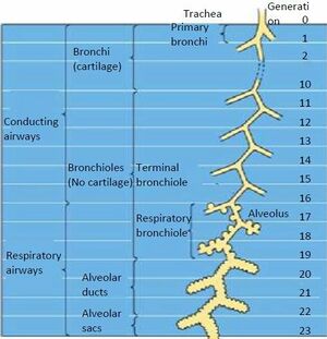
In all, there are approximately 23 generations (divisions) of airway in the human lung extending from trachea (generation 0) to the last order of terminal bronchioles (generation 23).
Trachea[edit | edit source]
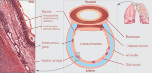
The trachea is a U-shaped structure that is composed of hyaline cartilage on the anterior and lateral walls, with the trachealis smooth muscle forming the posterior border of the trachea .Also the trachea is composed of several primary structural annular ligament.https://www.sciencedirect.com/topics/medicine-and-dentistry/trachealis-muscle , https://www.ncbi.nlm.nih.gov/books/NBK448070/. Posterior surface of the trachea is flat where its cartilaginous rings are incomplete and where it is related to the esophagus. (anatomy book )
- The trachea descends from the larynx into the lung.https://www.ncbi.nlm.nih.gov/books/NBK556044/#:~:text=The%20tracheobronchial%20tree%20is%20composed,the%20lungs%20for%20gas%20exchange.
- Is situated anterior to the oesophagus. (biblio)
- .Extends from cricoid cartilage to carina.https://www.ncbi.nlm.nih.gov/books/NBK556044/#:~:text=The%20tracheobronchial%20tree%20is%20composed,the%20lungs%20for%20gas%20exchange./2.0-2.5 cm in diameter.
- The trachea bifurcates into the right and left main bronchi , at the level of carina or sternum angle.https://www.ncbi.nlm.nih.gov/books/NBK448070/
- The tracheal diameter is about 24 to 26 mm in adult males and 22 to 24 mm in females10-12 cm long.
Trachea four layers[edit | edit source]
Inner mucosal layer = of ciliated pseudostratified columnar epithelium with goblet cells.
Submucosa is located just below the mucosa and contains nervous tissue and blood vessels.
The trachealis muscle found in the posterior wall of trachea.
Functions of trachea[edit | edit source]
The primary function of the trachea is to allow passage of inspired and expired air into and out of the lung.
Hyaline cartilage[edit | edit source]
- Series of 16 to 20 hyaline cartilage rings one top the other each is individually connected by an annular ligament.
- The tracheal cartilaginous rings, are responsible for keeping the trachea lumen open in spite of the changes in intrathoracic pressure that occur during respiration, and hence prevent air flow limitation.
Trachealis muscle[edit | edit source]
- Type of muscle = Smooth muscle
- Connects the ends of the C-shaped tracheal cartilages
- The trachealis muscle functions to constrict the airway by pulling the cartilages together, decrease the diameter , allowing for increased expiratory force during coughing with the smaller aperture allowing greater velocity to be achieved and the particle to be expelled. http://education.med.nyu.edu/Histology/courseware/modules/respiratory-sy/respiratory.07.html
This action is helpful, especially when eating food, which requires the expansion of the esophagus.
Bronchi[edit | edit source]
At the level of the fifth thoracic vertebra, the trachea divides into the left and right main bronchi. The right main bronchus has a length of 2.5 cm, and the left main bronchus is 5 cm long
Right main bronchi
- 2.5cm
- Wider
- More vertical.
- Left main bronchi
- 5cm
- Narrower
- More horizontal
Bronchioles[edit | edit source]
The bronchioles are the final air conductors, and by definition, lack cartilage altogether (and are therefore sometimes referred to as membranous) (Fig. 1.11). The bronchioles have no alveoli; alveoli are acquired more distally in the pulmonary acinus. The terminal bronchiole is the smallest conducting airway without alveoli in its walls. There are about 30,000 terminal bronchioles in the lungs, and each of these, in turn, directs air to approximately 10,000 alveoli. The cells that line the airways are columnar in shape and ciliated. Their nuclei are present at multiple levels in each cell—a phenomenon referred to as pseudostratification.
Resources[edit | edit source]
- bulleted list
- x
or
- numbered list
- x


