Sporting Hand and Wrist - What to Consider When Measuring Range of Motion: Difference between revisions
Carin Hunter (talk | contribs) No edit summary |
Carin Hunter (talk | contribs) No edit summary |
||
| Line 1: | Line 1: | ||
== Bony Anatomy == | == Bony Anatomy == | ||
The hand and wrist have a total of 27 bones arranged to [https://www.physio-pedia.com/Arthrokinematics roll, spin and slide] | The hand and wrist have a total of 27 bones arranged to [https://www.physio-pedia.com/Arthrokinematics roll, spin and slide]<ref>Maitland, G.D. Maitland's Peripheral Manipulations. 3rd Edition Edinburg: Elsevier Butterworth-Heinemann, 1999</ref>; allowing the hand to explore and control the environment and objects. | ||
The carpus is formed from eight small bones collectively referred to as the carpal bones. The carpal bones are bound in two groups of four bones: | The carpus is formed from eight small bones collectively referred to as the carpal bones. The carpal bones are bound in two groups of four bones: | ||
| Line 8: | Line 8: | ||
* the metacarpals – the five bones that comprise the middle part of the hand | * the metacarpals – the five bones that comprise the middle part of the hand | ||
* the phalanges (singular phalanx) – the 14 narrow bones that make up the fingers of each hand. Each finger has three phalanges (the distal, middle, and proximal); the thumb has two. | * the phalanges (singular phalanx) – the 14 narrow bones that make up the fingers of each hand. Each finger has three phalanges (the distal, middle, and proximal); the thumb has two. | ||
The hand is divided into three regions | The hand is divided into three regions<ref>Physical Examination of the Spine and Extremities. Hoppenfield, S. New York: Appleton-Century-Crofts, 1976.</ref> | ||
* Proximal region of the hand is the carpus (wrist) | * Proximal region of the hand is the carpus (wrist) | ||
* The middle region the metacarpus (palm) | * The middle region the metacarpus (palm) | ||
| Line 68: | Line 68: | ||
|} | |} | ||
Table summarises from - Kapandji I.A. The Physiology of the Joints: Volume 1, The Upper Limb. 5th Ed. London: Churchill Livingstone, 1982 | Table summarises from - Kapandji I.A. The Physiology of the Joints: Volume 1, The Upper Limb. 5th Ed. London: Churchill Livingstone, 1982 | ||
== Range of motion in the wrist and hand == | == Range of motion in the wrist and hand == | ||
{| class="wikitable" | {| class="wikitable" | ||
| Line 86: | Line 84: | ||
|2nd and 3rd metacarpals | |2nd and 3rd metacarpals | ||
|Median nerve (C6,7) | |Median nerve (C6,7) | ||
| | |60% midcarpal joint | ||
40% radiocarpal joint | |||
|- | |- | ||
| | | | ||
| Line 136: | Line 135: | ||
|2nd metacarpal | |2nd metacarpal | ||
|Radial nerve (C6,7) | |Radial nerve (C6,7) | ||
| | |40% midcarpal joint | ||
60% radiocarpal joint | |||
|- | |- | ||
| | | | ||
| Line 273: | Line 274: | ||
| | | | ||
|} | |} | ||
Table summarised from | Table summarised from Anatomy and Human Movement: Structure and Function. 6th Ed.<ref>Palastanga N, Soames R. Anatomy and Human Movement: Structure and Function. 6th Ed. London: Churchill Livingstone, 2012</ref> | ||
== Tools used to measure range of motion in the wrist and hand == | == Tools used to measure range of motion in the wrist and hand == | ||
| Line 299: | Line 300: | ||
== Considerations when measuring wrist flexion and extension == | == Considerations when measuring wrist flexion and extension == | ||
# Coupling | # Coupling | ||
<gallery widths="“250px”" heights="“350px”"> | Coupling occurs in the wrist while performing flexion–extension and radial–ulnar deviation movements. Maximal wrist range of motion is near the neutral position. The wrist should be placed at a neutral position in work station design to account for the naturally coupled wrist motion<ref>Li ZM, Kuxhaus L, Fisk JA, Christophel TH. Coupling between wrist flexion–extension and radial–ulnar deviation. Clinical biomechanics. 2005 Feb 1;20(2):177-83.</ref>.<gallery widths="“250px”" heights="“350px”"> | ||
File:E RD.png|Extension/radial deviation | File:E RD.png|Extension/radial deviation | ||
File:Neutral.png|Neutral | File:Neutral.png|Neutral | ||
Revision as of 08:43, 23 March 2021
Bony Anatomy[edit | edit source]
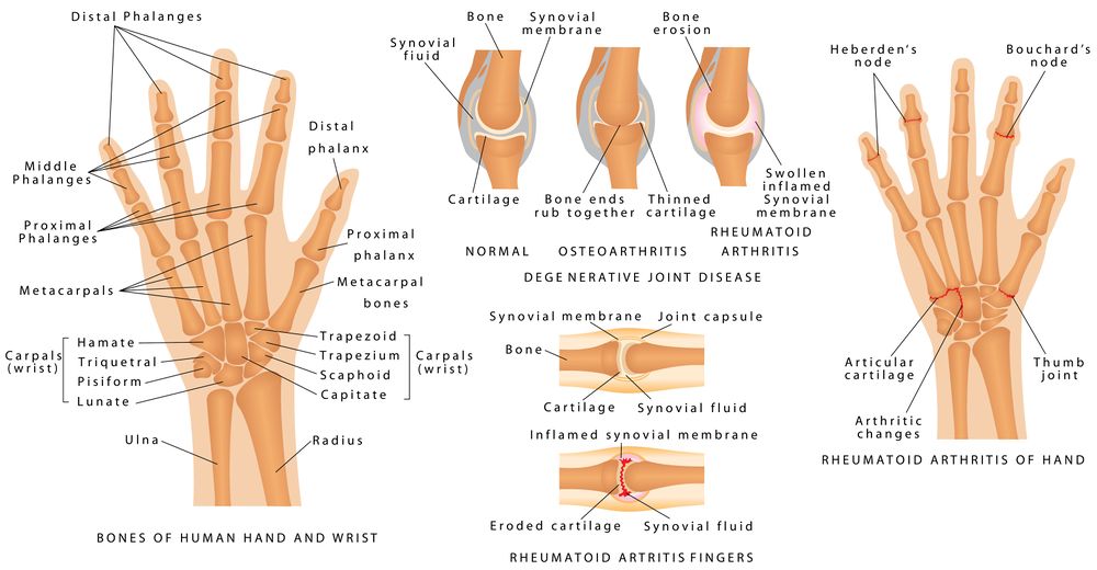
The hand and wrist have a total of 27 bones arranged to roll, spin and slide[1]; allowing the hand to explore and control the environment and objects.
The carpus is formed from eight small bones collectively referred to as the carpal bones. The carpal bones are bound in two groups of four bones:
- the pisiform, triquetrum, lunate and scaphoid on the upper end of the wrist
- the hamate, capitate, trapezoid and trapezium on the lower side of the hand.
Other bones of the hand are:
- the metacarpals – the five bones that comprise the middle part of the hand
- the phalanges (singular phalanx) – the 14 narrow bones that make up the fingers of each hand. Each finger has three phalanges (the distal, middle, and proximal); the thumb has two.
The hand is divided into three regions[2]
- Proximal region of the hand is the carpus (wrist)
- The middle region the metacarpus (palm)
- The distal region the phalanges (fingers)

Joints in the wrist and hand[edit | edit source]
| Joint | Proximal articulation | Distal Articulation | Type | Movement |
|---|---|---|---|---|
| Radiocarpal joint | Radius and articular disc/ concave | Scaphoid, lunate, triquetrum / – convex | Ellipsoid | Flexion-extension; Abduction-adduction |
| Midcarpal joint | Scaphoid. Lunate, Triquetrum | Trapezium, Trapezoid, Capitate, Hamate | Gliding | Flexion-extension; Abduction-adduction |
| Carpometacarpal joint (thumb) | 1st metacarpal | trapezium | Saddle | Flexion-extension; Abduction-adduction; circumduction; opposition |
| Carpometacarpal joint (fingers) | 2nd metacarpal
3rd metacarpal 4th metacarpal 5th metacarpal |
trapezoid, trapezium
capitate capitate, hamate hamate |
Ellipsoidal | Flexion-extension |
| Metacarpophalangeal joints | Metacarpals | phalangeal | Ellipsoidal | Flexion, extension, abduction, adduction, circumduction |
| Interphalangeal | Distal phlangeal | Proximal phalangeal | Hinge | Flexion (lots) Extension (minimal) |
Table summarises from - Kapandji I.A. The Physiology of the Joints: Volume 1, The Upper Limb. 5th Ed. London: Churchill Livingstone, 1982
Range of motion in the wrist and hand[edit | edit source]
| Movement | Muscles | Range | Origin | Insertion | Innervation | Joint |
| Flexion | Flexor carpi radialis | 0-80 | Medial Epicondyle of humerus | 2nd and 3rd metacarpals | Median nerve (C6,7) | 60% midcarpal joint
40% radiocarpal joint |
| Flexor carpi ulnaris | Medial epicondyle of humerus and sup.post. border of ulna | Pisiform, hamate and base 5th metacarpal | Ulnar nerve (C7,8) | |||
| Palmaris longus | Medial epicondyle of humerus | Flexor reinaculum and palmar aponeurosis | Median nerve (C8) | |||
| Flexor digitorum superficialis | Medial epicondyle of humerus, coronoid process of
ulna, and a ridge along lateral margin of anterior surface of radius |
Middle phalanges of each finger | Median nerve (C7,8 T1) | |||
| Flexor digitorum profundus | Anterior medial surface of body of ulna | Base of distal phalanx of thumb | Median nerve (C7,8, T1)
Ulnar nerve (C8, T1) |
|||
| Flexor pollicis longus | Anterior surface of radius and interosseous membrane | Base of distal phalanx of thumb | Median nerve (C8, T1) | |||
| Extension | Extensor carpi radialis longus | 0-70 | Lateral supracondylar ridge of humerus | 2nd metacarpal | Radial nerve (C6,7) | 40% midcarpal joint
60% radiocarpal joint |
| Extensor carpi radialis brevis | Lateral epicondyle of humerus | Distal and middle phalanges of each finger | Radial nerve (C6,7) | |||
| Extensor carpi ulnaris | Lateral epicondyle of humerus and posterior border of ulna | 5th metacarpal | Radial nerve (C7,8) | |||
| Extensor digitorum | Lateral epicondyle of humerus | Distal and middle phalanges of each finger | Radial nerve (C7,8) | |||
| Extensor indicis | Posterior surface of ulna | Tendon of extensor digitorum of index finger | Radial nerve (C7, 8) | |||
| Extensor digiti minimi | Lateral epicondyle of humerus | Tendon of extensor digitorum on 5th phalanx | Radial nerve (C7,8) | |||
| Extensor pollicis longus | Posterior surface of middle of radius and ulna and interosseous membrane | 1st metacarpal | Radial nerve (C7,8) | |||
| Extensor pollicis brevis | Posterior surface of middle of radius | Base of proximal phalanx of thumb | Radial nerve (C7,8) | |||
| Radial deviation | Flexor carpi radialias | 30 | Medial Epicondyle of humerus | 2nd and 3rd metacarpals | Median nerve (C6,7) | Radiocarpal Jt |
| Ulnar deviation | Flexor carpi ulnaris | 20 | Medial epicondyle of humerus and sup.post. border of ulna | Pisiform, hamate and base 5th metacarpal | Ulnar nerve (C7,8) | Radiocarpal Jt |
| Supination
(of forearm) |
Supinator | 60 | Lateral epicondyle of humerus and ridge near radial notch of ulna | Lateral surface of proximal one-third of radius | Radial nerve (C5,6) | Sup. and inf.radioulnar jt |
| Biceps brachii | Long head - supraglenoid tubercle; Short head - coracoid process of scapula | Radial tuberosity and bicipital aponeurosis | Musculocutaneous nerve (C5,6) | |||
| Brachioradialis | Medial and lateral borders of distal end of humerus | Superior to styloid process of radius | Radial Nerve (C5,6) | |||
| Pronation
(of forearm) |
Pronator teres | 40 | Medial epicondyle of humerus and coronoid process of ulna | Midlateral surface of radius | Median nerve (C6,7) | Sup.and inf.radioulnar jt |
| Pronator quadratus | Distal portion of shaft of ulna | Distal portion of shaft of radius | Median nerve (C8, T1) | |||
| Brachioradialis | Medial and lateral borders of distal end of humerus | Superior to styloid process of radius | Radial nerve (C5,6) | |||
| Distal Interphalangeal | ||||||
| Proximal Interphalangeal |
Table summarised from Anatomy and Human Movement: Structure and Function. 6th Ed.[3]
Tools used to measure range of motion in the wrist and hand[edit | edit source]

Goniometer position for wrist flexion and extension

Inclinometer

Cellphone Inclinometer


| Goniometer | Inclinometer | Cellphone Inclinometer | |
|---|---|---|---|
| Pros | Compact, easy to use and portable | Accurate and measures multiple planes | Accurate, measures multiple planes, free downloadable appconvenient |
| Limitations | Only one plane of movement | Bulky and expensive |
Considerations when measuring wrist flexion and extension[edit | edit source]
- Coupling
Coupling occurs in the wrist while performing flexion–extension and radial–ulnar deviation movements. Maximal wrist range of motion is near the neutral position. The wrist should be placed at a neutral position in work station design to account for the naturally coupled wrist motion[4].

Extension/radial deviation

Neutral

Flexion/Ulnar deviation


2. Open versus closed hand

Flexion
- Open hand = joint
- Closed hand = soft tissue
Extension
- Open hand = soft tissue
- Closed hand = joint
Reason to measure[edit | edit source]
The main reason to measure is that you have an accurate baseline to base your rehab and progression off.
- ↑ Maitland, G.D. Maitland's Peripheral Manipulations. 3rd Edition Edinburg: Elsevier Butterworth-Heinemann, 1999
- ↑ Physical Examination of the Spine and Extremities. Hoppenfield, S. New York: Appleton-Century-Crofts, 1976.
- ↑ Palastanga N, Soames R. Anatomy and Human Movement: Structure and Function. 6th Ed. London: Churchill Livingstone, 2012
- ↑ Li ZM, Kuxhaus L, Fisk JA, Christophel TH. Coupling between wrist flexion–extension and radial–ulnar deviation. Clinical biomechanics. 2005 Feb 1;20(2):177-83.

