Thumb Ligament Injuries: Difference between revisions
No edit summary |
No edit summary |
||
| Line 1: | Line 1: | ||
== INTRODUCTION == | <div class="noeditbox">This article or area is currently under construction and may only be partially complete. Please come back soon to see the finished work! ({{REVISIONDAY}}/{{REVISIONMONTH}}/{{REVISIONYEAR}})</div> | ||
== INTRODUCTION == <div class="editorbox"> '''Original Editor '''- [[User:User Name|User Name]] <br> | |||
'''Top Contributors''' - {{Special:Contributors/{{FULLPAGENAME}}}}</div> | |||
== Clinically Relevant Anatomy<br> == | |||
add text here relating to '''''clinically relevant''''' anatomy of the condition<br> | |||
== Mechanism of Injury / Pathological Process<br> == | |||
add text here relating to the mechanism of injury and/or pathology of the condition<br> | |||
== Clinical Presentation == | |||
add text here relating to the clinical presentation of the condition<br> | |||
== Diagnostic Procedures == | |||
add text here relating to diagnostic tests for the condition<br> | |||
== Outcome Measures == | |||
add links to outcome measures here (see [[Outcome Measures|Outcome Measures Database]]) | |||
== Management / Interventions<br> == | |||
add text here relating to management approaches to the condition<br> | |||
== Differential Diagnosis<br> == | |||
add text here relating to the differential diagnosis of this condition<br> | |||
== Resources <br> == | |||
add appropriate resources here | |||
== References == | |||
<references />. | |||
The Metacarpophalangeal (MCP) joint of the thumb are stabilize by two major ligaments. The ulnar collateral ligament (UCL) and the Radial collateral ligament (RCL) . The UCL is more commonly injured, usually from forced radial deviation (abduction) of the thumb, while the RCL are rarely injuried. However, in severe type of injuries, both ligaments may be ruptured.<ref>Weiss L, Weiss J, Pobre T. Oxford American handbook of physical medicine & rehabilitation. Oxford University Press, USA; 2010 Mar 15.</ref> | The Metacarpophalangeal (MCP) joint of the thumb are stabilize by two major ligaments. The ulnar collateral ligament (UCL) and the Radial collateral ligament (RCL) . The UCL is more commonly injured, usually from forced radial deviation (abduction) of the thumb, while the RCL are rarely injuried. However, in severe type of injuries, both ligaments may be ruptured.<ref>Weiss L, Weiss J, Pobre T. Oxford American handbook of physical medicine & rehabilitation. Oxford University Press, USA; 2010 Mar 15.</ref> | ||
Revision as of 13:09, 29 October 2020
== INTRODUCTION ==
Clinically Relevant Anatomy
[edit | edit source]
add text here relating to clinically relevant anatomy of the condition
Mechanism of Injury / Pathological Process
[edit | edit source]
add text here relating to the mechanism of injury and/or pathology of the condition
Clinical Presentation[edit | edit source]
add text here relating to the clinical presentation of the condition
Diagnostic Procedures[edit | edit source]
add text here relating to diagnostic tests for the condition
Outcome Measures[edit | edit source]
add links to outcome measures here (see Outcome Measures Database)
Management / Interventions
[edit | edit source]
add text here relating to management approaches to the condition
Differential Diagnosis
[edit | edit source]
add text here relating to the differential diagnosis of this condition
Resources
[edit | edit source]
add appropriate resources here
References[edit | edit source]
. The Metacarpophalangeal (MCP) joint of the thumb are stabilize by two major ligaments. The ulnar collateral ligament (UCL) and the Radial collateral ligament (RCL) . The UCL is more commonly injured, usually from forced radial deviation (abduction) of the thumb, while the RCL are rarely injuried. However, in severe type of injuries, both ligaments may be ruptured.[1]
Clinically Relevant Anatomy[edit | edit source]
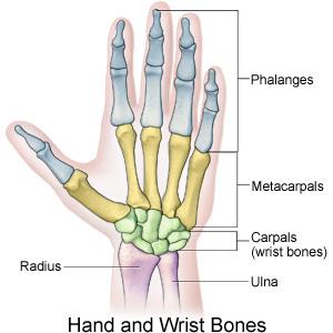
The thumb MCP is similar in anatomical appearance to those of the finger,but essentially functions as a hinge or ginglymus joints. The articular morphology found in this joint makes it the most varied motion of all joints, with range of motion of 6 to 86 degree in flexion-extension. [2]
- flexor pollicis brevis (FBP)
- Abductor pollicis brevis (APB) muscles insert partially on the sesamoids and provide stability against hyperextension forces.
The ligamentous anatomy is analogous to that seen in the finger MCP joints, with extrinsic tendons providing additional support
Physical exam/Evaluation[edit | edit source]
History taking, including mechanism, finger position during injury, the presence of deformity, previous treatment received, and subjective sense of stability of the injured thumb.
• Neurovascular exam must determine motor function, perfusion, and
sensation.
• Weakness with pinch function usually exists in ligament ruptures.
• Examine the base of the thumb for ligamentous laxity and compare it
to the uninjured hand:
• Examine the thumb with 20–30* of fl exion.
• Carefully abduct the thumb passively and compare the angle of
deviation to the uninjured thumb.
• An angulation of >30* on the injured thumb or >15* compared to
the uninjured thumb is diagnostic for a ligamentous injury.
• Radiographs should be obtained to assess for the presence of a Stener
lesion or a fracture fragment.
•further assessment of joint should be perfomed using a digital block may be necessary to complete a full examination
because of pain and swelling in the acute setting
Differential diagnosis[edit | edit source]
• First metacarpal or proximal phalanx fractures
• First CMC joint arthritis
• Volar plate injury


