Ultrasound Scans: Difference between revisions
Kim Jackson (talk | contribs) No edit summary |
Kim Jackson (talk | contribs) (Formatting) |
||
| Line 15: | Line 15: | ||
The uses of ultrasound imaging are broad, important uses include | The uses of ultrasound imaging are broad, important uses include | ||
* | * Muscles and tendons | ||
* | * Abdominal organs | ||
* | * Heart | ||
* | * Breast | ||
* | * Arteries and veins | ||
=== Advantages === | === Advantages === | ||
While it may provide less anatomical detail than techniques such as CT or MRI, it has several advantages which make it ideal in numerous situations | While it may provide less anatomical detail than techniques such as CT or MRI, it has several advantages which make it ideal in numerous situations | ||
* Studies the function of moving structures in real-time | |||
Studies the function of moving structures in real-time | * Emits no ionizing radiation | ||
* Contains speckle that can be used in elastography | |||
Emits no ionizing radiation | * It is very safe to use and does not appear to cause any adverse effects, although information on this is not well documented | ||
* It is also relatively inexpensive and quick to perform. | |||
Contains speckle that can be used in elastography | * Ultrasound scanners can be taken to critically ill patients in intensive care units, avoiding the danger caused while moving the patient to the radiology department | ||
* The real time moving image obtained can be used to guide drainage and biopsy procedures | |||
It is very safe to use and does not appear to cause any adverse effects, although information on this is not well documented | * Doppler capabilities on modern scanners allow the blood flow in arteries and veins to be assessed. | ||
It is also relatively inexpensive and quick to perform. | |||
Ultrasound scanners can be taken to critically ill patients in intensive care units, avoiding the danger caused while moving the patient to the radiology department | |||
The real time moving image obtained can be used to guide drainage and biopsy procedures | |||
Doppler capabilities on modern scanners allow the blood flow in arteries and veins to be assessed. | |||
This short video gives a animated insight into how US scans work | This short video gives a animated insight into how US scans work | ||
{{#ev:youtube|https://www.youtube.com/watch?v=I1Bdp2tMFsY|width}}<ref>NIBIB gov How Ultrasound Works Available from: https://www.youtube.com/watch?v=I1Bdp2tMFsY (last accessed 22.9.2019)</ref> | {{#ev:youtube|https://www.youtube.com/watch?v=I1Bdp2tMFsY|width}}<ref>NIBIB gov How Ultrasound Works Available from: https://www.youtube.com/watch?v=I1Bdp2tMFsY (last accessed 22.9.2019)</ref> | ||
| Line 46: | Line 37: | ||
The ability to make correct ultrasonographic diagnosis in sports injuries is improving as advancing technology allows for high-resolution images in contemporary medical ultrasound. Ultrasonography demonstrates tissue structure with two-dimensional grayscale images. Blood flow in the tissue can be rapidly depicted with colour and power Doppler technique. Ultrasonography is the preferred imaging modality to study soft tissue lesions dynamically. With high-resolution images possible with ultrasonography, injuries of the muscle, tendon, ligament, bursa, bony structure, cartilage, and subcutaneous tissue can be accurately diagnosed (if the examiner is well trained). Recently, compact ultrasound machine machines are becoming increasingly available, leading to prompt ultrasonographic diagnosis of sports injuries on the field<ref>Chiang YP, Wang TG, Hsieh SF. [https://www.sciencedirect.com/science/article/pii/S092964411300009X Application of ultrasound in sports injury.] Journal of Medical Ultrasound. 2013 Mar 1;21(1):1-8. Available from: https://www.sciencedirect.com/science/article/pii/S092964411300009X (last accessed 22.9.2019)</ref>. | The ability to make correct ultrasonographic diagnosis in sports injuries is improving as advancing technology allows for high-resolution images in contemporary medical ultrasound. Ultrasonography demonstrates tissue structure with two-dimensional grayscale images. Blood flow in the tissue can be rapidly depicted with colour and power Doppler technique. Ultrasonography is the preferred imaging modality to study soft tissue lesions dynamically. With high-resolution images possible with ultrasonography, injuries of the muscle, tendon, ligament, bursa, bony structure, cartilage, and subcutaneous tissue can be accurately diagnosed (if the examiner is well trained). Recently, compact ultrasound machine machines are becoming increasingly available, leading to prompt ultrasonographic diagnosis of sports injuries on the field<ref>Chiang YP, Wang TG, Hsieh SF. [https://www.sciencedirect.com/science/article/pii/S092964411300009X Application of ultrasound in sports injury.] Journal of Medical Ultrasound. 2013 Mar 1;21(1):1-8. Available from: https://www.sciencedirect.com/science/article/pii/S092964411300009X (last accessed 22.9.2019)</ref>. | ||
== References == | |||
[[Category:Assessment]] | [[Category:Assessment]] | ||
[[Category:Extended_Scope]] | [[Category:Extended_Scope]] | ||
Revision as of 17:49, 31 October 2019
Ultrasound[edit | edit source]
Medical ultrasonography uses high frequency broadband sound waves in the megahertz range that are reflected by tissue to varying degrees to produce (up to 3D) images (commonly associated with imaging the fetus in pregnant women). Ultrasonography is generally considered safe imaging with the World Health Organizations saying:
"Diagnostic ultrasound is recognized as a safe, effective, and highly flexible imaging modality capable of providing clinically relevant information about most parts of the body in a rapid and cost-effective fashion"[1].
The uses of ultrasound imaging are broad, important uses include
- Muscles and tendons
- Abdominal organs
- Heart
- Breast
- Arteries and veins
Advantages[edit | edit source]
While it may provide less anatomical detail than techniques such as CT or MRI, it has several advantages which make it ideal in numerous situations
- Studies the function of moving structures in real-time
- Emits no ionizing radiation
- Contains speckle that can be used in elastography
- It is very safe to use and does not appear to cause any adverse effects, although information on this is not well documented
- It is also relatively inexpensive and quick to perform.
- Ultrasound scanners can be taken to critically ill patients in intensive care units, avoiding the danger caused while moving the patient to the radiology department
- The real time moving image obtained can be used to guide drainage and biopsy procedures
- Doppler capabilities on modern scanners allow the blood flow in arteries and veins to be assessed.
This short video gives a animated insight into how US scans work
Application of Ultrasound in Sports Injury[edit | edit source]
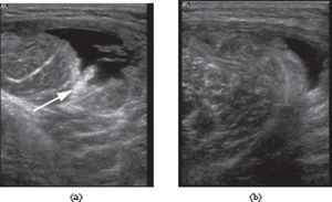
The ability to make correct ultrasonographic diagnosis in sports injuries is improving as advancing technology allows for high-resolution images in contemporary medical ultrasound. Ultrasonography demonstrates tissue structure with two-dimensional grayscale images. Blood flow in the tissue can be rapidly depicted with colour and power Doppler technique. Ultrasonography is the preferred imaging modality to study soft tissue lesions dynamically. With high-resolution images possible with ultrasonography, injuries of the muscle, tendon, ligament, bursa, bony structure, cartilage, and subcutaneous tissue can be accurately diagnosed (if the examiner is well trained). Recently, compact ultrasound machine machines are becoming increasingly available, leading to prompt ultrasonographic diagnosis of sports injuries on the field[3].
References[edit | edit source]
- ↑ WHO study group. TRAINING IN DIAGNOSTIC ULTRASOUND: ESSENTIALS, PRINCIPLES AND STANDARDS.Spain 1998 Available from: https://apps.who.int/iris/bitstream/handle/10665/42093/WHO_TRS_875.pdf;jsessionid=992840A3C30619985E80DCD5243B24CA?sequence=1 (last accessed 22.9.2019).
- ↑ NIBIB gov How Ultrasound Works Available from: https://www.youtube.com/watch?v=I1Bdp2tMFsY (last accessed 22.9.2019)
- ↑ Chiang YP, Wang TG, Hsieh SF. Application of ultrasound in sports injury. Journal of Medical Ultrasound. 2013 Mar 1;21(1):1-8. Available from: https://www.sciencedirect.com/science/article/pii/S092964411300009X (last accessed 22.9.2019)


