Management of Thoracic Outlet Syndrome: Difference between revisions

mNo edit summary
mNo edit summary
 
(27 intermediate revisions by 7 users not shown)
Line 1: Line 1:
<div class="editorbox">
'''Original Editors''' - [[User:Chelsey Walker|Chelsey Walker]], [[User:Jacqueline Keller|Jacqueline Keller]], [[User:Katie Schwarz|Katie Schwarz]], [[User:Jenny Nordin|Jenny Nordin]], [[User:Chris Slininger|Chris Slininger]] as part of the [[Texas State University Evidence-based Practice Project]]


'''Top Contributors''' - {{Special:Contributors/{{FULLPAGENAME}}}}
== Introduction ==
</div>
[[Thoracic Outlet Syndrome (TOS)|Thoracic outlet syndrome]] (TOS) is characterized by the compression of neurovascular structures as they pass through the thoracic outlet, leading to a spectrum of symptoms ranging from mild pain to limb-threatening complications. It remains a complex and controversial condition in musculoskeletal medicine and rehabilitation. The syndrome encompasses various anatomical structures and can present with diverse clinical manifestations.


== Definition/Description  ==
Clinically relevant anatomy highlights the intricate passageways through which neurovascular structures traverse, including the interscalene, costoclavicular, and subcoracoid spaces. Anatomical abnormalities and acquired conditions contribute to the compression of these structures, leading to the development of TOS.


The term ‘thoracic outlet syndrome’ describes compression of the neurovascular structures as they exit through the thoracic outlet (cervicothoracobrachial region). The thoracic outlet is marked by the anterior scalene muscle anteriorly, the middle scalene posteriorly, and the first rib inferiorly.<ref name=":0">Jacky Laulan et al. Thoracic outlet syndrome: definition, aetiological factors, diagnosis, management and occupational impact. J Occup Rehabil. 2011 Sep; 21(3): 366–373.&nbsp;</ref> <ref name=":1">Gul Koknel Talu. Thoracic outlet syndrome. 2005; 17(2)</ref>[25][26]
Epidemiologically, TOS affects approximately 8% of the population, with a higher prevalence in women due to anatomical and physiological factors. It predominantly affects individuals between the ages of 20 and 50, with the brachial plexus being the most commonly affected structure.


This condition has emerged as one of the most controversial topics in musculoskeletal medicine and rehabilitation<ref name=":2">Hooper T, Denton J, McGalliard M, Brismée J, Sizer P. Thoracic outlet syndrome: a controversial clinical condition. Part 1: anatomy, and clinical examination/diagnosis. Journal Of Manual Manipulative Therapy (Maney Publishing) [serial online]. June 2010;18(2):74-83. Available from: CINAHL Plus with Full Text, Ipswich, MA. Accessed November 20, 2011. </ref> [2]. This controversy extends to almost every aspect of the pathology including the definition, incidence, pathoanatomical contributions, diagnosis, and treatment.  
The diagnosis of TOS relies on a comprehensive evaluation, including history, physical examination, and provocative tests. Differential diagnosis is essential to distinguish TOS from other pathologies with similar presentations. Imaging studies and electrodiagnostic evaluation are valuable adjuncts in confirming the diagnosis.


The term ‘TOS’ does not specify the structure being compressed. Investigators namely identify two main categories of TOS: the vascular form (arterial or venous), which raises few diagnostic problems, and the neurological form, which occurs in more than 95-99% of all cases of TOS. Therefore the syndrome should be differentiated by using the terms arterial TOS (ATOS), venous TOS (VTOS) or neurogenic (NTOS).<ref name=":0" /><ref name=":1" /> [25][26]
Treatment approaches for TOS encompass both non-surgical and surgical interventions, tailored to the individual patient's presentation and severity of symptoms. Physical therapy, including stretching, strengthening, and postural re-education, plays a crucial role in managing TOS-related symptoms.
{{#ev:youtube|H_tGstO-0N4}}


== Clinically Relevant Anatomy  ==
== Medical Management ==


The first narrowing area is the most proximal and is named the interscalene triangle: This triangle is bordered by the anterior scalene muscle anteriorly, the middle scalene muscle posteriorly, and the medial surface of the first rib inferiorly. The presence of the scalene minimus muscle and the fact that both the anterior and middle scalene muscles have their insertion in the first rib (which can cause overlapping) can cause a narrow space and therefore compression . The brachial plexus and the subclavian artery pass through this space.  
* Nonsteroidal anti-inflammatory drugs have been prescribed to reduce pain and inflammation.
* Botulinum injections to the anterior and middle scalenes have also been found to temporarily reduce pain and spasm from neurovascular compression, further research is needed because there are discrepancies in the literature<ref>Jordan SE, Ahn SS, Freischlag JA, Gelabert HA, Machleder HI. Selective botulinum chemodenervation of the scalene muscles for treatment of neurogenic thoracic outlet syndrome. Annals of vascular surgery. 2000 Jul 1;14(4):365-9.</ref><ref>Christo PJ, Christo DK, Carinci AJ, Freischlag JA. Single CT-guided chemodenervation of the anterior scalene muscle with botulinum toxin for neurogenic thoracic outlet syndrome. Pain Medicine. 2010 Apr 1;11(4):504-11.</ref>.<ref>Finlayson HC, O’Connor RJ, Brasher PM, Travlos A. Botulinum toxin injection for management of thoracic outlet syndrome: a double-blind, randomized, controlled trial. Pain. 2011 Sep 1;152(9):2023-8.</ref>
* Surgical management of TOS should only be considered after conservative treatment has been proven ineffective.<ref name=":0">Ciampi P, Scotti C, Gerevini S, De Cobelli F, Chiesa R, Fraschini G, Peretti GM. Surgical treatment of thoracic outlet syndrome in young adults: single centre experience with minimum three-year follow-up. International orthopaedics. 2011 Aug;35(8):1179-86.</ref>. However, limb-threatening complications of vascular TOS have been indicated for surgical intervention.<ref name=":1">Hooper TL, Denton J, McGalliard MK, Brismée JM, Sizer Jr PS. Thoracic outlet syndrome: a controversial clinical condition. Part 2: non-surgical and surgical management. Journal of Manual & Manipulative Therapy. 2010 Sep 1;18(3):132-8.</ref>
* Surgery to treat thoracic outlet syndrome may be performed using several different approaches, including: transaxillary approach, supraclavicular approach and infraclavicular approach.<ref name=":0" />


The second passageway is called the costoclavicular triangle which is bordered anteriorly by the middle third of the clavicle, posteromedially by the first rib, and posterolaterally by the upper border of the scapula. The subclavian vein, artery and plexus brachialis crosses this costoclavicular region and then further enters the subcoracoïd space. Just distal to the insterscalene triangle. Compression of these structures can occur as a result of congenital abnormalities, trauma to the first rib or clavicle, and structural changes in the subclavian muscle or the costocoracoid ligament.  
* '''Transaxillary approach.'''


The last passageway is called the subcoracoid or sub-pectoralis minor space: This last passageway is beneath the coracoid process just under the pectoralis minor tendon. The borders of the thoraco-coraco-pectoral space include the coracoid process superiorly, the pec minor anteriorly, and ribs 2-4 posteriorly. Shortening of the Pectoralis Major can lead to a narrowing of this last space and therefore compression of the neurovascular structures during hyperabduction.<ref name=":1" /><ref name=":3">Laulan J, Fouquet B, Rodaix C, Jauffret P, Roquelaure Y, Descatha A. Thoracic Outlet Syndrome: Definition, Aetiological Factors, Diagnosis, Management and Occupational Impact. Journal Of Occupational Rehabilitation [serial online]. September 2011;21(3):366-373. Available from: CINAHL Plus with Full Text, Ipswich, MA. Accessed November 7, 2011.</ref><ref name=":4">César Fernàndez et al.; Manual Therapy for Musculoskeletal Pain Syndromes; Elsevier, 2016&nbsp;</ref> [3][24][26]
The first rib forms the common denominator for all causes of nerve and artery compression in this region, so that its removal generally improves symptoms. The surgeon makes an incision in the chest to access the first rib, divides the muscles in front of the rib, and removes a portion of the first rib to relieve compression, without disturbing the nerves or blood vessels<ref name=":2">Urschel Jr HC, Razzuk MA. Neurovascular compression in the thoracic outlet: changing management over 50 years. Annals of surgery. 1998 Oct;228(4):609</ref><ref name=":3">Siracuse JJ, Johnston PC, Jones DW, Gill HL, Connolly PH, Meltzer AJ, Schneider DB. Infraclavicular first rib resection for the treatment of acute venous thoracic outlet syndrome. Journal of Vascular Surgery: Venous and Lymphatic Disorders. 2015 Oct 1;3(4):397-400.</ref>.<ref name=":0" />


Certain anatomical abnormalities can be potentially compromising to the thoracic outlet as well. These include the presence of a cervical rib, congenital soft tissue abnormalities, clavicular hypomobility <ref name=":2" />[2], and functionally acquired anatomical changes<ref name=":3" /> [3].&nbsp;Soft tissue abnormalities may create compression or tension loading of the neurovascular structures found within the thoracic outlet (such as hypertrophy , a broader middle scalene attachment on the 1st rib or fibrous bands that increase the stiffness,…).<br>
* '''Supraclavicular approach'''


[[Image:Tos.jpg]]
it has been advocated to perform first rib resection and scalenectomy, a safe and effective procedure, characterized by a shorter operative time and a complication rate lower or comparable to that of transaxillary first rib resection.
This approach repairs compressed blood vessels. The surgeon makes an incision just under the neck to expose the brachial plexus region. Then he looks for signs of trauma or muscles contributing to compression near the first rib. The first rib may be removed if necessary to relieve compression<ref name=":3" /><ref name=":2" />.<ref name=":0" />


== Epidemiology/Etiology  ==
* '''Infraclavicular approach.'''


TOS affects approximately 8% of the population and is 3-4 times as frequent In woman as in men between the age of 20 and 50 years. Females have less-developed muscles, a greater tendency for drooping shoulders owing to additional breast tissue, a narrowed thoracic outlet and an anatomical lower sternum, these factors change the angle between the scalene muscles and consequently cause a higher prevalence in women.<ref name=":3" /><ref name=":4" /><ref>Lindgren K. Thoracic outlet syndrome. International Musculoskeletal Medicine. March 2010;32(1):17-24. Accessed November 2, 2011.</ref>[3][16] [24] The mean age of people effected with TOS is 30s-40s; it is rarely seen in children. Almost all cases of TOS (95-98%) affect the brachial plexus; the other 2-5% affecting vascular structures, such as the subclavian artery and vein.&nbsp;
In this approach, the surgeon makes an incision under the collarbone and across the chest. This procedure may be used to treat compressed veins that require extensive repair.<ref name=":0" /><ref name=":3" />


There are several factors which can cause TOS: Cervical ribs are present in approximately 0.5-0.6% of the population, 50-80% of which are bilateral, and 10-20% produce symptoms; the female to male ratio is 2:1. Cervical ribs and the fibromuscular bands connected to them are the cause of most neural compression.<ref>Boezaart, AP, et al. Neurogenic thoracic outlet sndrome: A case report and review of the literature. International Journal of Shoulder Surgery. 2010;4:27-35.</ref> [8] Fibrous bands are a more common cause of TOS than rib anomalies.
* '''Neurogenic TOS'''


Congenital factors: 
Surgical decompression should be considered for those with true neurological signs or symptoms. These include weakness, wasting of the hand intrinsic muscles, and conduction velocity less than 60 m/sec. The first rib can be a major contributor to TOS. There is controversy, however, regarding the necessity of a complete resection to reduce the chance of reattachment of the scalenes, scar tissue development, or bony growth of the remaining tissue. In addition to the first rib, cervical ribs are removed, scalenectomies can be performed, and fibrous bands can be excised<ref name=":1" />. Terzis found that the supraclavicular approach to treatment was an effective and precise surgical method<ref>Terzis JK, Kokkalis ZT. Supraclavicular approach for thoracic outlet syndrome. Hand. 2010 Sep 1;5(3):326-37.</ref>
* Cervical rib<ref name=":5">Thoracic outlet syndrome: anatomy. Erdogan Atasoy, MD. Hand Clin 20 (2004) 7–14
</ref><ref name=":6">Diagnosis of thoracic outlet syndrome. Richard J. Sanders MD, Sharon L. Hammond MD and Neal M. Rao BA. J Vasc Surg. 2007 Sep;46(3):601-4.</ref><ref>Harold C. Urschel, Jr., MD. Transaxillary First Rib Resection for Thoracic Outlet Syndrome.  Operative techniques in thoracic and cardiovascular surgery. 2005 </ref>[4][9][10] 
* Prolonged transverse process
* Anomalous muscles 
* Fibrous anomalies (transversocostal, costocostal)
* Abnormalities of the insertion of the scalene muscles<ref name=":5" /> [4]
* Fibrous muscular bands<ref name=":5" />[4] 
* Exostosis of the first rib 
* Cervicodorsal scoliosis<ref name=":7">Michael Hamm, LMP. Impact of massage therapy in the treatment of linked pathologies: Scoliosis, costovertebral dysfunction, and thoracic outlet syndrome.Journal of Bodywork and Movement Therapies (2006) 10, 12–20. </ref>[11] 
* Congenital uni- or bilateral elevated scapula 
* Location of the A. or V. Subclavian in relation to the M. scalene anterior
Acquired conditions: 
* Postural factors
* Dropped shoulder condition<ref name=":5" />[4][12] 
* Wrong work posture (standing or sitting without paying attention to the physiological curvature of the spine) 
* Heavy mammaries 
* Trauma<ref name=":7" />[11] 
* Clavicle fracture<ref name=":5" />[4] 
* Rib fracture<ref name=":5" />[4] 
* Hyperextension neck injury, whiplash<ref name=":6" /><ref name=":8">Andrew K Chang, MD et al.; Thoracic Outlet Syndrome in Emergency Medicine; Medscape, Mar 04, 2014</ref>[6][9] 
* Repetitive stress injuries (repetitive injury most often form sitting at a keyboard for long hours)<ref name=":8" />[6]<br>
Muscular causes:
* Hypertrophy of the scalene muscles 
* Decrease of the tonus of the M. trapezius, M. levator scapulae, M.rhomboids 
* Shortening of the scalene muscles, M. trapezius, M. levator scapulae, pectoral muscles <br><br>  


== Characteristics/Clinical Presentation  ==
* '''Arterial TOS'''


Signs and symptoms of thoracic outlet syndrome vary from patient to patient due to the location of nerve and/or vessel involvement. Symptoms range from mild pain and sensory changes to limb threatening complications in severe cases.  
Decompression can include cervical and/or first rib removal and scalene muscle revision. The subclavian can then be inspected for degeneration, dilation, or aneurysm. Saphenous vein graft or synthetic prosthesis can then be used if necessary<ref name=":1" />


Patients with thoracic outlet syndrome will most likely present pain anywhere between the neck, face and occipital region or into the chest, shoulder and upper extremity and paresthesia in upper extremity. The patient may also complain of altered or absent sensation, weakness, fatigue, a feeling of heaviness in the arm and hand. The skin can also be blotchy or discolored. A different temperature can also be observed.
* '''Venous TOS'''


Signs and symptoms are typically worse when the arm is abducted overhead and externally rotated with the head rotated to the same or the opposite side. As a result activities such as overhead throwing, serving a tennis ball, painting a ceiling, driving, or typing may exacerbate symptoms.<ref>Sandra J. Shultz et al.; Examination of Musculoskeletal Injuries; Human kinetics, 2010&nbsp;
Thrombolytic therapy is the first line of treatment for these patients. Because of the risk of recurrence, many recommend that removal of the first rib is necessary even when thrombolytic therapy completely opens the vein. The results of a study show that the infraclavicular approach is a safe and effective treatment for acute VTOS. They had no brachial plexus or phrenic nerve injuries<ref name=":4">Schneider DB, Dimuzio PJ, Martin ND, Gordon RL, Wilson MW, Laberge JM, Kerlan RK, Eichler CM, Messina LM. Combination treatment of venous thoracic outlet syndrome: open surgical decompression and intraoperative angioplasty. Journal of vascular surgery. 2004 Oct 1;40(4):599-603.</ref>.<ref name=":3" /> Angioplasty can then be used to treat those with venous stenosis<ref name=":1" />
</ref><ref>Jacky Laulan et al. Thoracic outlet syndrome: definition, aetiological factors, diagnosis, management and occupational impact. J Occup Rehabil. 2011 Sep; 21(3): 366–373.</ref> [21][23]


When the upper plexus (C5,6,7) is involved there is pain in the side of the neck and this pain may radiate to the ear and face. Often the pain radiates from the ear posteriorly to the rhomboids and anteriorly over the clavicle and pectoralis regions. The pain may move laterally down the radial nerve area. Headaches are not uncommon when the upper plexus is involved.  
In venous or arterial TOS, medication can be administered to dissolve blood clots prior to thoracic outlet compression. It may also be necessary to conduct a procedure to remove a clot from the vein or artery or repair the vein or artery prior to thoracic outlet decompression.<ref name=":4" />


Patients with lower plexus (C8,T1) involvement typically have symptoms which are present in the anterior and posterior shoulder region and radiate down the ulnar side of the forearm into the hand, the ring and small fingers.<ref name=":2" /><ref name=":4" /> [24][2]
Some larger-chested women have sagging shoulders that increase pressure on the neurovascular structures in the thoracic outlet. A supportive bra with wide and posterior-crossing straps can help reduce tension. Extreme cases may resort to breast-reduction surgery to relieve TOS and other biomechanical problems<ref name=":5">Vanti C, Natalini L, Romeo A, Tosarelli D, Pillastrini P. Conservative treatment of thoracic outlet syndrome. Eura medicophys. 2007 Mar 1;43:55-70.</ref><ref>Buckley L, Schub E. Thoracic Outlet Syndrome. October 2010; Accessed November 2,2011</ref>.<ref>Boezaart AP, Haller A, Laduzenski S, Koyyalamudi VB, Ihnatsenka B, Wright T. Neurogenic thoracic outlet syndrome: A case report and review of the literature. International journal of shoulder surgery. 2010 Apr;4(2):27.</ref>


There are four categories of thoracic outlet syndrome and each presents with unique signs and symptoms (see Table 1). Typically TOS does not follow a dermatomal or myotomal pattern unless there is nerve root involvement, which will be important in determining your PT diagnosis and planning your treatment <ref name=":2" /><ref name=":3" />[2][3]. <br>  
== Physical Therapy Management ==
Conservative management should be the first strategy to treat TOS since this seems to be effective at decreasing symptoms, facilitating return to work, and improving function, but yet a few studies have evaluated the optimal exercise program as well as the difference between conservative management and no treatment. <ref name=":6">Vanti C. et al., Conservative treatment of thoracic outlet syndrome A review of the literature, Europa Medicophysica, 2006, Volume 42</ref>


Conservative management includes physical therapy, which focuses mainly on patient education, pain control, range of motion, nerve gliding techniques, strengthening and stretching<ref>Crosby CA, Wehbé MA. Conservative treatment for thoracic outlet syndrome. Hand clinics. 2004 Feb 1;20(1):43-9.</ref>.


{| width="700" cellspacing="1" cellpadding="1" border="1" align="center"
=== Stage 1 ===
|-
The aim of the initial stage is to decrease the patient’s symptoms. This may be achieved by patient education, in which TOS, bad postures, the prognosis and the importance of therapy compliance are explained. Furthermore, some patients who sleep with the arms in an overhead, abducted position should get some information about their sleeping posture to avoid waking up at night. These patients should sleep on their uninvolved side or supine, potentially by pinning down the sleeves. The Cyriax release test may be used if a ‘release phenomenon’ is present. This technique completely unloads the neurovascular structures in the thoracic outlet before going to bed.<ref name=":1" />
| '''Arterial TOS'''
| '''Venous TOS'''
| '''True TOS'''
| '''Disputed Neurogenic TOS'''
|-
|
*Young adult with&nbsp;vigorous arm&nbsp;activity
*Pain in the hand
*Claudication
*Pallor
*Cold intolerance
*Paresthesias
*S/s usually appear spontaneously


|
=== Cyriax Release Maneuver ===
*Younger men with vigorous arm activity
*Cyanosis
*Feeling of heaviness
*Paresthesia in fingers and hand (result of edema)
*Edema of the arm&nbsp;


|
* Elbows flexed to 90°
*Hx of neck trauma
* Towels create a passive shoulder girdle elevation
*Pain, paresthesia, numbness, and/or weakness
* Supported spine and the head in neutral
*Occipital headaches
* The position is held until peripheral symptoms are produced. The patient is encouraged to allow symptoms to occur as long as can be tolerated for up to 30 minutes, observing for a symptom decrescendo as time passes.<ref name=":1" />
*S/s present day and/or night
*Loss of fine motor skills
*Cold intolerance (possible Raynaud's phenomenon)
*Objective weakness
*Compressors*: s/s day&gt;night


|
*Hx of neck trauma
*Pain, paresthesia, and "feeling" of weakness
*Occipital headaches
*Nocturnal paresthesias that often wake patient
*Loss of fine motor skills
*Cold intolerance (possible Raynaud's phenomenon)
*Subjective weakness
*Releasers*: s/s night&gt;day


|}
The patient’s breathing techniques need to be evaluated as the scalenes and other accessory muscles often compensate to elevate the ribcage during inspiration. Encouraging diaphragmatic breathing will lessen the workload on already overused or tight scalenes and can possibly reduce symptoms.<ref name=":1" />


Compressors* - a patient that experiences symptoms throughout the daytime while using prolonged postures resulting in increased tension or compression of the thoracic outlet
=== Scapula Settings and Control ===
In the treatment, you first have to start with scapula settings and control.


Releasers* - a patient that experiences a release phenomenon (release of tension or compression to thoracic outlet) that often awakes them at night
This is important to establishing normal scapula muscle recruitment and control in the resting position. Once this is achieved then the program is progressed to maintaining scapula control while both motion and load are applied. The programme begins in lower ranges of abduction and is gradually progressed further up into abduction and flexion range until muscles are being retrained in functional movement patterns at higher ranges of elevation.


== Differential Diagnosis  ==
=== Control the Humeral Head Position ===
It is also important to control the humeral head position. Specific drills are given to facilitate humeral head control. The most common aberrant position of the humeral head is an increase in anterior placement of the humeral head. A useful strategy to help facilitate co-contraction of the rotator cuff to help stabilize and centralize the humeral head is to facilitate a mid-level isometric contraction of the rotator cuff by applying resistance to the humeral head (Dark et al., 2007).


Due the it's variability, TOS can be difficult to tease out from other pathologies with similar presentations. A thorough history and evaluation must be done to determine if the patient’s symptoms are truly TOS. The following pathologies are common differential diagnosis for TOS [13][14]: <br>• Carpal tunnel syndrome <br>• De Quervain’s tenosynovitis <br>• Lateral epicondylitis <br>• Medial epicondylitis <br>• Complex regional pain syndrome (CRPS I or II). <br>• Horner’s Syndrome <br>• Raynaud’s disease <br>• Cervical disease (especially discogenic) <br>• Brachial plexus trauma <br>• Systemic disorders: inflammatory disease, esophageal or cardiac disease <br>• Upper extremity deep venous thrombosis (UEDVT), Paget-Schroetter syndrome <br>• Rotator cuff pathology <br>• Glenohumeral joint instability <br>• Nerve root involvement <br>• Malignancies (local tumours)<br>• Chest pain, angina<br>• Vasculitis<br>• Thoracic 4 syndrome<br>• Sympathetic-mediated pain
Further on in the treatment, this may be integrated into movement patterns. First in slow controlled concentric/eccentric motion drills, later isolated muscle strengthening drills.


<br>Systematic causes of brachial plexus pain include: <br>• Pancoast’s Syndrome <br>• Radiation induced brachial plexopathy <br>• Parsonage Turner Syndrome [2][3]
=== Serratus Anterior Recruitment and Control ===
Abduction external rotation strategies described above are often sufficient to trigger serratus anterior recruitment and control without the risk of over-activating pectoral minor muscle


There are conditions that can coexist with TOS. It is important to identify these conditions because they should be treated separately. These associated conditions include: <br>• carpal tunnel syndrome <br>• peripheral neuropathies (like ulnar nerve entrapment at the elbow, shoulder tendinitis and impingement syndrome) <br>• fibromyalgia of the shoulder and neck muscles <br>• cervical disc disease (like cervical spondylosis and herniated cervical disk) <br><br>
=== Stage 2 ===
Once the patient has control over his/her symptoms, the patient can move to this stage of treatment. The goal of this stage is to directly address the tissues that create structural limitations of motion and compression. How this should be done is one of the most discussed topics of this pathology. Some examples of methods that are used in the literature are.


== Outcome Measures  ==
* Massage
* Strengthening of the levator scapulae, sternocleidomastoid and upper trapezius (This group of muscles open the thoracic outlet by raising the shoulder girdle and opening the costoclavicular space)
* Stretching of the pectoralis, lower trapezius and scalene muscles (These muscles close the thoracic outlet)
* Postural correction exercises
* Relaxation of shortened muscles<ref name=":5" />
* Aerobic exercises in a daily home exercise program<ref name=":7">Lindgren KA. Conservative treatment of thoracic outlet syndrome: a 2-year follow-up. Archives of physical medicine and rehabilitation. 1997 Apr 1;78(4):373-8.</ref><ref name=":5" />


[[DASH Outcome Measure|DASH (Disability of Arm Shoulder and Hand)]]
=== Exercises ===


SPADI (Shoulder Pain And Disability Index)  
# Shoulder exercises to restore the range of motion and so provide more space for the neurovascular structures.                  Exercise: Lift your shoulders backwards and up, flex your upper thoracic spine and move the shoulders forward and down. Then straighten the back and repeat 5 to 10 times.
# ROM of the upper cervical spine.                                                                                                                                                                            Exercise: Lower your chin 5 to 10 times against your chest, while you are standing with the back of your head against a wall. The effectiveness of this exercise can be enlarged by pressing the head down by hands.
# Activation of the scalene muscles is the most important exercises. These exercises help to normalize the function of the thoracic aperture as well as all the malfunctions of the first rib. Exercises are Anterior scalene (Press your forehead 5 times against the palm of your hand for a duration of 5 seconds, without creating any movement), Middle scalene (Press your head sidewards against your palm), Posterior scalene (Press your head backwards against your palm
# Stretching exercises


NPRS (Numeric Pain Rating Scale)
=== Other Interventions ===


[[Short-form McGill Pain Questionnaire|McGill Pain Questionnaire]]<ref name="Vanti" />  
* Repositioning/mobilization of the shoulder girdle and pelvis joints: cervicothoracic, sternoclavicular, acromioclavicular, and costotransverse joints<ref name=":1" /> <ref name=":5" />
* Glenohumeral mobilizations in end-range elevation with the elbow supported in extension<ref name=":7" />
* Taping: some patients with severe symptoms respond to additional taping, adhesive bandages or braces that elevate or retract the shoulder girdle.<ref name=":8">Vanti C, Natalini L, Romeo A, Tosarelli D, Pillastrini P. Conservative treatment of thoracic outlet syndrome. Eura medicophys. 2007 Mar 1;43:55-70.</ref><ref name=":6" />


<br>  
=== Manipulative Treatment to Mobilize the First Rib ===
These should be carried out with caution and only after a thorough assessment as they can provoke irritation and pain symptoms in some patients<ref name=":8" />


== Diagnostic Procedures&nbsp;  ==
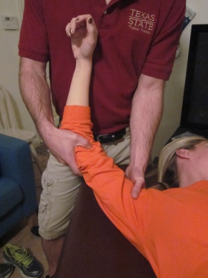
# Posterior Glenohumeral Glide with Arm Flexion:                                                                                                                                                    The patient is supine. The mobilization hand contacts the proximal humerus avoiding corocoid process. The force is directed posterolaterally (direction of thumb).
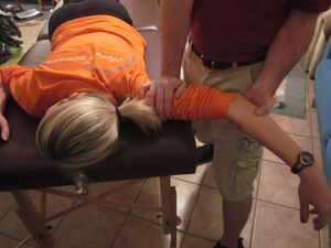
# Anterior Glenohumeral Glide with Arm Scaption:                                                                                                                                                  The patient is prone. The mobilization hand contacts the proximal humerus avoiding acromion process. The force is directed anteromedially.
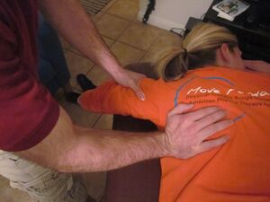
# Inferior Glenohumeral Glide:                                                                                                                                                                                            The patient is prone. The stabilizing hand holds the proximal humerus, the humerus distal to the lateral acromion process. The mobilization hand contacts the axillary border of the scapula. Mobilize the scapula in a craniomedial direction along the ribcage.<ref name=":1" />


The diagnosis of TOS is essentially based on history, physical examination, provocative tests, and if needed ultrasound, radiological evaluation and electrodiagnostic evaluation. It must always kept in mind that TOS diagnosis is usually confirmed by elimination of other causes with similar clinical presentation. Especially differential diagnosis of cervical radiculopathies and upper extremity entrapment neuropathies can be hard (McGillicuddy 2004). [26][28] In order to diagnose accurately, the clinical presentation must be evaluated as either neurogenic (compression of the brachial plexus) or vascular (compression of the subclavian vessels). TOS manifestations are varied and there is no single definitive test, which makes it difficult to diagnose. [21][24]  
[[File:Firstribselfmob.jpg|thumb|[[/www.physio-pedia.com/File:Firstribselfmob.jpg|Firstribselfmob.jpg]]]]


== Examination<br>  ==
'''First Rib Mobilization''': Patient seated. Thin sheet strap positioned around the first rib. Pull strap towards the opposite hip. Neck retracted, contralateral lateral flexion, and ipsilateral rotation. Ipsilateral head rotation emphasizes scalene stretch. Contralateral rotation emphasizes rib mobilization.


The following includes common examination findings seen with TOS that should be evaluated; however, this is not an all-inclusive list and examination should be individualized to the patient.  
[[File:Posteriorghglidearmflex.jpg|thumb|[[/www.physio-pedia.com/File:Posteriorghglidearmflex.jpg|Posteriorghglidearmflex.jpg]]]]
 '''Posterior Glenohumeral Glide with Arm Flexion:''' Patient supine. Mobilizing hand contacts proximal humerus avoiding corocoid process. Force is directed posterolaterally (direction of thumb).


<br>'''History'''[15]


*Make sure to take a thorough history, clear any red flags, and ask the patient how signs/symptoms have affected his/her function.


- Type of symptoms


- Location and amplitude of symptoms


- Irritability of symptoms


- Onset and development over time


- Aggravating/alleviating factors
[[File:Antghglidearmscaption.JPG|thumb|[[/www.physio-pedia.com/File:Antghglidearmscaption.JPG|Antghglidearmscaption.JPG]]]]
 '''Anterior Glenohumeral Glide with Arm Scaption:''' Patient prone. Mobilizing hand contacts proximal humerus avoiding acromion process. Force is anteromedially.


- Disability


'''Physical Examination[15][16]'''


*Observation


- Posture <br>- Cyanosis <br>- Edema <br>- Paleness <br>- Atrophy


<br>
'''Inferior Glenohumeral Glide:''' Patient prone. Stabilizing hand holds proximal humerus. Mobilizing hand contacts axillary border of scapula. Mobilize scapula in craniomedial direction along ribcage.
[[File:Infghglide.JPG|thumb|[[/www.physio-pedia.com/File:Infghglide.JPG|Infghglide.JPG]]]]
===Post-Op Physical Therapy===


*Palpation


- Temperature changes <br>- Supraclavicular fossa<br>- Scalene muscles (tenderness)<br>- Trapezius muscle (tenderness)<br>


*Neurological Screen
If a patient does require surgery, then physical therapy should follow immediately to prevent scar tissue and return the patient to full function.
*MMT &amp; Flexibility of following muscles:


- Scalene <br>- Pectoralis major/minor <br>- Levator scapulae <br>- Sternocleidomastoid <br>- Serratus anterior


'''<br>Special Tests[2][16] '''


*[http://www.physio-pedia.com/Roos_Stress_Test Elevated Arm Stress/ Roos test]: the patient has arms at 90° abduction and the therapist puts downwards pressure on the scapula as the patient opens and closes the fingers. If the TOS symptoms are reproduced within 90 seconds, the test is positive. [24]
*[http://www.physio-pedia.com/Adsons_Test Adson's]: the patient is asked to rotate the head and elevate the chin toward the affected side. If the radial pulse on the side is absent or decreased then the test is positive, showing the vascular component of the neurovascular bundle is compressed by the scalene muscle or cervical rib. [24]
*Wright's: the patient’s arm is hyper abducted. If there is a decrease or absence of a pulse on one side then the test is positive, showing the axillary artery is compressed by the pectoralis minor muscle or coracoid process due to stretching of the neurovascular bundle. [24]
*Cyriax Release: the patient is seated or standing. The examiner stands behind patient and grasps under the forearms, holding the elbows at 80 degrees of flexion with the forearms and wrists in neutral. The examiner leans the patient’s trunk posteriorly and passively elevated the shoulder girdle. This position is held for up to 3 minutes. The test is positive when paresthesia and/or numbness (release phenomenon) occurs, including reproduction of symptoms. (Hooper et. al., 2010 &amp; Brismee et. al., 2004)
*Supraclavicular Pressure: the patient is seated with the arms at the side. The examiner places his fingers on the upper trapezius and thumb on the anterior scalene muscle near the first rib. Then the examiner squeezes the fingers and thumb together for 30 seconds. If there is a reproduction of pain or paresthesia the test is positive, this addresses compromise to brachial plexus through scalene triangles. (Hooper et al., 2010)
*Costoclavicular Maneuver: this test may be used for both neurological and vascular compromise. The patient brings his shoulders posteriorly and hyperflexes his chin. A decrease in symptoms means that the test is positive and that he neurogenic component of the neurovascular bundle is compressed. [24]
*Upper Limb Tension: These tests are designed to put stress on neurological structures of upper limb. The shoulder, elbow, forearm, wrist and fingers are kept in specific position to put stress on particular nerve (nerve bias) and further modification in position of each joint is done as "sensitizer". (http://www.physio-pedia.com/Neurodynamic_Assessment)
*Cervical Rotation Lateral Flexion: The test is performed with the patient in sitting. The cervical spine is passively and maximally rotated away from the side being tested. While maintaining this position, the spine is gently flexed as far as possible moving the ear toward the chest. A test is considered positive when the lateral flexion movement is blocked.


<br>


{| cellspacing="1" cellpadding="1" border="1" width="500"
|-
| <span style="font-size: 13.28px;">Test</span>
| <span style="font-size: 13.28px;">Sensitivity</span>
| <span style="font-size: 13.28px;">Specificity</span>
| <span style="font-size: 13.28px;">LR+</span>
| <span style="font-size: 13.28px;">LR-</span>
|-
| <span style="font-size: 13.28px; line-height: 19.92px;">Elevated Arm Stress</span>
| <span style="font-size: 13.28px; line-height: 19.92px;">52-84%&nbsp;</span>
| <span style="font-size: 13.28px; line-height: 19.92px;">30-100%&nbsp;</span>
| <span style="font-size: 13.28px; line-height: 19.92px;">1.2-5.2&nbsp;</span>
| <span style="font-size: 13.28px; line-height: 19.92px;">0.4-0.53&nbsp;</span>
|-
| <span style="font-size: 13.28px; line-height: 19.92px;">Adson's</span>
| <span style="font-size: 13.28px; line-height: 19.92px;">79%</span>
| <span style="font-size: 13.28px; line-height: 19.92px;">74-100%</span>
| <span style="font-size: 13.28px; line-height: 19.92px;">3.29</span>
| <span style="font-size: 13.28px; line-height: 19.92px;">0.28&nbsp;</span>
|-
|
<span style="font-size: 13.28px; line-height: 19.92px;">Wright's</span>


| <span style="font-size: 13.28px; line-height: 19.92px;">70-90%</span>
| <span style="font-size: 13.28px; line-height: 19.92px;">29-53%</span>
| <span style="font-size: 13.28px; line-height: 19.92px;">1.27-1.49</span>
| <span style="font-size: 13.28px; line-height: 19.92px;">0.34-0.57&nbsp;</span>
|-
| <span style="font-size: 13.28px; line-height: 19.92px;">Cyriax Release</span>
| <span style="font-size: 13.28px; line-height: 19.92px;">NT&nbsp;</span>
| <span style="font-size: 13.28px; line-height: 19.92px;">77-97%</span>
| <span style="font-size: 13.28px; line-height: 19.92px;">NA</span>
| <span style="font-size: 13.28px; line-height: 19.92px;">NA&nbsp;</span>
|-
| <span style="font-size: 13.28px; line-height: 19.92px;">Supraclavicular Pressure</span>
| <span style="font-size: 13.28px; line-height: 19.92px;">NT&nbsp;</span>
| <span style="font-size: 13.28px; line-height: 19.92px;">85-98%</span>
| <span style="font-size: 13.28px; line-height: 19.92px;">NA</span>
| <span style="font-size: 13.28px; line-height: 19.92px;">NA&nbsp;</span>
|-
| <span style="font-size: 13.28px; line-height: 19.92px;">Costoclavicular Maneuver</span>
| <span style="font-size: 13.28px; line-height: 19.92px;">NT</span>
| <span style="font-size: 13.28px; line-height: 19.92px;">53-100%</span>
| <span style="font-size: 13.28px; line-height: 19.92px;">NA</span>
| <span style="font-size: 13.28px; line-height: 19.92px;">NA&nbsp;</span>
|-
| <span style="font-size: 13.28px; line-height: 19.92px;">Upper Limb Tension</span>
| <span style="font-size: 13.28px; line-height: 19.92px;">90%</span>
| <span style="font-size: 13.28px; line-height: 19.92px;">38%</span>
| <span style="font-size: 13.28px; line-height: 19.92px;">1.5</span>
| <span style="font-size: 13.28px; line-height: 19.92px;">&nbsp;0.3&nbsp;</span>
|-
| <span style="font-size: 13.28px; line-height: 19.92px;">Cervical Rotation Lateral Flexion</span>
| <span style="font-size: 13.28px; line-height: 19.92px;">100%</span>
| NT
| NA
| NA
|}


<br>
== Conclusion ==
Thoracic outlet syndrome (TOS) presents a complex challenge, involving compression of neurovascular structures in the thoracic outlet. It demands a comprehensive approach to diagnosis and management, encompassing both conservative and surgical interventions. Physical therapy plays a crucial role in symptom management and rehabilitation, focusing on patient education, posture correction, strengthening, and stretching exercises. Surgical options, when necessary, aim to alleviate compression and restore function. Postoperative physical therapy is essential for optimizing recovery and preventing complications. Ongoing research and multidisciplinary collaboration are vital for advancing our understanding and treatment of this condition.


*Electrodiagnostic evaluation and imaging
== References ==
 
<references />
Nerve conduction studies and electromyography are often helpful as components of the diagnostic evaluation of patients with suspected TOS. Nerve conduction studies usually reveal decreased ulnar sensorial potentials, decreased median action potentials, normal or close to normal ulnar motor and median sensorial potentials (Huang and Zager 2004). Vascular TOS can be identified with venography and arteriography. <br>Besides the electrophysiological studies, imaging studies can provide useful information in the diagnosis of TOS. Cervical spine and chest x-rays are important in the identification of bony abnormalities (such as cervical ribs or a “peaked C7 transverse processes)<br><br>
[[Category:Rehabilitation Protocols]]
 
[[Category:Neurology]]
== Medical Management <br>  ==
 
Nonsteroidal anti-inflammatory drugs have been prescribed to reduce pain and inflammation. Botulinum injections to the anterior and middle scalenes have also found to temporarily reduce pain and spasm from neurovascular compression, further research is needed because there are discrepancies in the literature. [29] Level of evidence 1B, [30] Level of evidence 2B, [31] Level of evidence 4. Surgical management of TOS should only be considered after conservative treatment has been proven ineffective. [32] Level of evidence 2A<br>However, limb-threatening complications of vascular TOS have been indicated for surgical intervention. [17] Level of evidence 2B<br>Surgery to treat thoracic outlet syndrome, may be performed using several different approaches, including: transaxillary approach, supraclavicular approach and infraclavicular approach. [32] Level of evidence 2A
 
<br>
 
*Transaxillary approach. The first rib forms the common denominator for all causes of nerve and artery compression in this region, so that its removal generally improves symptoms. Surgeon makes an incision in the chest to access the first rib, divide the muscles in front of the rib and remove a portion of the first rib to relieve compression, without disturbing the nerves or blood vessels.[32] Level of evidence 2A, [33] Level of evidence 4, [34] Level of evidence 3A
*Supraclavicular approach has been advocated to perform first rib resection and scalenectomy, a safe and effective procedure, characterized by a shorter operative time and having a complication rate lower or comparable to that of transaxillary first rib resection. <br>This approach repairs compressed blood vessels. The surgeon makes an incision just under the neck to expose the brachial plexus region. Then he looks for signs of trauma or muscles contributing to compression near the first rib. The first rib may be removed if necessary to relieve compression. [32] Level of evidence 2A, [33] Level of evidence 4, [34] Level of evidence 3A
*Infraclavicular approach. In this approach, the surgeon makes an incision under the collarbone and across the chest. This procedure may be used to treat compressed veins that require extensive repair. [32] Level of evidence 2A, [34] Level of evidence 3A
*Neurogenic TOS: Surgical decompression should be considered for those with true neurological signs or symptoms. These include weakness, wasting of the hand intrinsic muscles, and conduction velocity less than 60 m/sec. The first rib can be a major contributor to TOS. There is controversy, however, regarding the necessity of a complete resection to reduce the chance of reattachment of the scalenes, scar tissue development, or bony growth of the remaining tissue. In addition to the first rib, cervical ribs are removed, scalenectomies can be performed, and fibrous bands can be excised[17] Level of evidence 2B. Terzis found that the supraclavicular approach to treatment to be an effective and precise surgical method [18] Level of evidence 3B.
*Arterial TOS: Decompression can include cervical and/or first rib removal and scalene muscle revision. The subclavian can then be inspected for degeneration, dilation, or aneurysm. Saphenous vein graft or synthetic prosthesis can then be used if necessary[17] Level of evidence 2B.
*Venous TOS: Thrombolytic therapy is the first line of treatment for these patients. Because of the risk of recurrence, many recommend removal of the first rib is necessary even when thrombolytic therapy completely opened the vein. The results of a study show that infraclavicular approach is a safe and effective treatment for acute VTOS. They had no brachial plexus or phrenic nerve injuries. [33] Level of evidence 4,[35] Level of evidence 2A<br>Angioplasty can then be used to treat those with venous stenosis[17] Level of evidence 2B.
 
In venous or arterial TOS, medication can be administered to dissolve blood clots prior to thoracic outlet compression. It may also be to conduct a procedure to remove a clot from the vein or artery or repair the vein or artery prior to thoracic outlet decompression. [35] Level of evidence 2A
 
Some larger-chested women have sagging shoulders that increase pressure on the neurovascular structures in the thoracic outlet. A supportive bra with wide and posterior-crossing straps can help reduce tension. Extreme cases may resort to breast-reduction surgery to relieve TOS and other biomechanical problems.[15]Level of evidence 1C, [8] Level of evidence 4, [14] <br><br>
 
== Physical Therapy Management <br>  ==
 
Conservative management should be the first strategy to treat TOS since this seems to be effective at decreasing symptoms, facilitating return to work and improving function, but yet a few studies have evaluated the optimal exercise program as well as the difference between a conservative management and no treatment.[36] Level of evidence 1A Conservative management includes physical therapy, which focuses mainly on patient education, paincontrol, range of motion, nerve gliding techniques, strengthening and stretching. [37] Level of evidence 2A&nbsp;
 
<br>Stage 1:  
 
&nbsp;The aim of the initial stage is to decrease the patient’s symptoms. This may be achieved by patient education, in which TOS, bad postures, the prognosis and the importance of therapy compliance are explained. Furthermore some patients who sleep with the arms in an overhead, abducted position should get some information about their sleeping posture to avoid waking up at night. These patients should sleep on their uninvolved side or in supine, potentially by pinning down the sleeves. The Cyriax release test may be used if a ‘release phenomenon’ is present. This technique completely unloads the neurovascular structures in the thoracic outlet before going to bed. Also cervical traction in combination with a hot pack and light exercise may reduce pain and irritable symptoms for some acute patients. [17] Level of evidence 2B<br>
 
<br>
 
Cyriax release maneuver<br>- Elbows flexed to 90° <br>- Towels create a passive shoulder girdle elevation <br>- Supported spine and the head in neutral <br>- The position is held until peripheral symptoms are produced. The patient is encouraged to allow symptoms to occur as long as can be tolerated up to 30 minutes, observing for a symptom decrescendo as time passes. [17] Level of evidence 2B
 
<br>The patient’s breathing techniques need to be evaluated as the scalenes and other accessory muscles often compensate to elevate the ribcage during inspiration. Encouraging diaphragmatic breathing will lessen the work load on already overused or tight scalenes and can possibly reduce symptoms. [17] Level of evidence 2B
 
<br>Stage 2: <br>Once the patient has control over his/her symptoms, the patient can move to this stage of treatment. The goal of this stage is to directly address the tissues that create structural limitations of motion and compression. How this should be done is one of the most discussed topics of this pathology. Some examples of methods that are used in the literature are.
 
<br>• Massage <br>• Strengthening of the levator scapulae, sternocleidomastoid and upper trapezius <br>= This group of muscles open the thoracic outlet by raising the shoulder girdle and opening the costoclavicular space<br>• Stretching of the pectoralis, lower trapezius and scalene muscles <br>= These muscles close the thoracic outlet<br>• Postural correction exercises <br>• Relaxation of shortened muscles [36] Level of evidence 1A<br>• Aerobic exercises in a daily home exercise program: [36] Level of evidence 1A, [38] Level of evidence 2B
 
<br>
 
1) Shoulder exercises to restore the range of motion and so provide more space for the neurovascular structures.<br>Exercise: Lift your shoulders backwards and up, flex your upper thoracic spine and move the shoulders forward and down. Then straightened the back and repeat 5 to 10 times.
 
<br>2) ROM of the upper cervical spine <br>Exercise: Lower your chin 5 to 10 times against your chest, while you are standing with the back of your head against a wall. The effectiveness of this exercise can be enlarged by pressing the head down by hands.
 
<br>3) Activation of the scalene muscles are the most important exercises. These exercises help to normalize the function of the thoracic aperture as well as all the malfunctions of the first rib. <br>Exercises: <br>- Anterior scalene: Press your forehead 5 times against the palm of your hand for a duration of 5 seconds, without creating any movement. <br>- Middle scalene: Press your head sidewards against your palm.<br>- Posterior scalene: Press your head backwards against your palm.
 
<br>4) Stretching exercises<br>• Taping: some patients with severe symptoms respond to additional taping, adhesive bandages or braces that elevate or retract the shoulder girdle.[15] Level of evidence 1C, [36] Level of evidence 1A<br>• Manipulative treatment to mobilize the first rib<br> Could provoke irritation and pain symptoms in some patients [36] Level of evidence 1A<br>
 
• Repositioning/mobilization of the shoulder girdle and pelvis joints: cervicothoracic, sternoclavicular, acromionclavicular, and costotransverse joints [36] Level of evidence 1A, [17] Level of evidence 2B<br>• Glenohumeral mobilizations in end-range elevation with the elbow supported in extension [38] Level of evidence 2B
 
<br>
 
A) Posterior Glenohumeral Glide with Arm Flexion: <br>The patient is supine. The mobilizisation hand contacts the proximal humerus avoiding corocoid process. The force is directed posterolaterally (direction of thumb).
 
<br>B) Anterior Glenohumeral Glide with Arm Scaption: <br>The patient is prone. The mobilization hand contacts the proximal humerus avoiding acromion process. The force is directed anteromedially.
 
<br>C) Inferior Glenohumeral Glide: <br>The patient is prone. The stabilizing hand holds the proximal humerus the humerus distal to the lateral acromion process. The mobilization hand contacts the axillary border of the scapula. Mobilize the scapula in a craniomedial direction along the ribcage. [17] Level of evidence 2B
 
<br>
 
[[Image:Firstribselfmob.jpg|200x300px]]'''&nbsp;First Rib Mobilization:''' Patient seated. Thin sheet strap positioned around first rib. Pull strap towards opposite hip. Neck retracted, contralateral lateral flexion, and ipsilateral rotation. Ipsilateral head rotation emphasizes scalene stretch. Contralateral rotation emphasizes rib mobilization.
 
[[Image:Posteriorghglidearmflex.jpg|200x300px]]&nbsp;'''Posterior Glenohumeral Glide with Arm Flexion: '''Patient supine. Mobilizing hand contacts proximal humerus avoiding corocoid process. Force is directed posterolaterally (direction of thumb).
 
[[Image:Antghglidearmscaption.JPG|300x200px]]&nbsp;'''Anterior Glenohumeral Glide with Arm Scaption:''' Patient prone. Mobilizing hand contacts proximal humerus avoiding acromion process. Force is anteromedially.<br>
 
[[Image:Infghglide.JPG|300x200px]]&nbsp;'''Inferior Glenohumeral Glide: '''Patient prone. Stabilizing hand holds proximal humerus. Mobilizing hand contacts axillary border of scapula. Mobilize scapula in craniomedial direction along ribcage.
 
<br>
 
<u>Post-Op Physical Therapy<ref name="godges" /></u><br>If a patient does require surgery, then physical therapy should follow immediately to prevent scar tissue and return the patient to full function.
 
<br><br>
 
== Key Research  ==
 
Hooper T, Denton J, McGalliard M, Brismée J, Sizer P. Thoracic outlet syndrome: a controversial clinical condition. Part 1: anatomy, and clinical examination/diagnosis. Journal Of Manual and Manipulative Therapy.&nbsp;June 2010;18(2):74-83.<br>
 
Hooper T, Denton J, McGalliard M, Brismée J, Sizer P. Thoracic outlet syndrome: a controversial clinical condition. Part 2: non-surgical and surgical management. Journal of Manual and Manipulative Therapy.&nbsp;June 2010;18(3):132-138.
 
== Resources <br>  ==
 
[http://thoracicoutletsyndromes.com/index.html thoracicoutletsyndromes.com]
 
[http://www.ninds.nih.gov/disorders/thoracic/thoracic.htm NINDS Thoracic Outlet Syndrome Information Page]
 
== Clinical Bottom Line  ==
 
TOS can present in numerous ways due to the variety of tissues that can be involved (arteries, veins, nervous, and muscular tissue) and the different anatomical sites in which compression or entrapment can occur. In general, treatment for TOS should initially begin conservatively according to a literature review by Vanti et al, however, firm conclusions cannot be drawn from this review due to the lack of high quality evidence <ref name="Vanti" />. Conservative treatment seems to be effective at reducing symptoms, improving function, and facilitating return to work when compared to surgery.6 Higher quality studies are needed to compare conservative treatment to surgery, and even no treatment at all <ref name="Vanti" />. Physical therapy can assist patients given a TOS diagnosis utilizing an impairment-based approach, addressing muscle imbalances and postural changes that these patients commonly present with. <br>
 
== Presentations  ==
<div class="coursebox">
{| width="100%" cellspacing="4" cellpadding="4" border="0" class="FCK__ShowTableBorders"
|-
| align="center" | <imagemap>
Image:TOS part 1 presentation title.png|200px|border|left|
rect 0 0 830 452 [https://www.youtube.com/watch?v=2NCO4lzIN30]
desc none
</imagemap>
| [https://www.youtube.com/watch?v=2NCO4lzIN30/ '''Conservative Management of Thoracic Outlet Syndrome Part 1''']  
This presentation was created by Walt Lingerfelt, Fellow in training at Evidence in Motion.
 
[https://www.youtube.com/watch?v=2NCO4lzIN30/ Conservative Management of Thoracic Outlet Syndrome Part 1 / View the presentation]
 
|}
</div> <div class="coursebox">
{| width="100%" cellspacing="4" cellpadding="4" border="0" class="FCK__ShowTableBorders"
|-
| align="center" | <imagemap>
Image:TOS part 2 presentation title.png|200px|border|left|
rect 0 0 830 452 [https://www.youtube.com/watch?v=iVzx-XH1gwE]
desc none
</imagemap>
| [https://www.youtube.com/watch?v=iVzx-XH1gwE/ '''Conservative Management of Thoracic Outlet Syndrome Part 2''']  
This presentation was created by Walt Lingerfelt, Fellow in training at Evidence in Motion.
 
[https://www.youtube.com/watch?v=iVzx-XH1gwE/ Conservative Management of Thoracic Outlet Syndrome Part 2 / View the presentation]
 
|}
</div>
== References  ==
 
1. ↑ Christo P, McGreevy K. Updated perspectives on neurogenic thoracic outlet syndrome. Current Pain And Headache Reports [serial online]. February 2011;15(1):14-21. Available from: MEDLINE with Full Text, Ipswich, MA. Accessed November 27, 2011( Level of evidence: 1A )
 
<br>2. ↑ 2.0 2.1 2.2 2.3 2.4 2.5 2.6 Hooper T, Denton J, McGalliard M, Brismée J, Sizer P. Thoracic outlet syndrome: a controversial clinical condition. Part 1: anatomy, and clinical examination/diagnosis. Journal Of Manual Manipulative Therapy (Maney Publishing) [serial online]. June 2010;18(2):74-83. Available from: CINAHL Plus with Full Text, Ipswich, MA. Accessed November 20, 2011. (Level of evidence: 1B)
 
<br>3. ↑ 3.0 3.1 3.2 3.3 3.4 Laulan J, Fouquet B, Rodaix C, Jauffret P, Roquelaure Y, Descatha A. Thoracic Outlet Syndrome: Definition, Aetiological Factors, Diagnosis, Management and Occupational Impact. Journal Of Occupational Rehabilitation [serial online]. September 2011;21(3):366-373. Available from: CINAHL Plus with Full Text, Ipswich, MA. Accessed November 7, 2011.(Level of evidence: C)
 
<br>4. ↑ 4.0 4.1 4.2 4.3 4.4 4.5 4.6 Thoracic outlet syndrome: anatomy. Erdogan Atasoy, MD. Hand Clin 20 (2004) 7–14 (Level of evidence: B)
 
<br>5. ↑ Surgery of peripheral nerves: a case-based approach. Rajiv Midha, Eric L. Zager. 2008 - 288 pages. (Level of evidence: C)
 
<br>6. ↑ 6.0 6.1 6.2 Andrew K Chang, MD et al.; Thoracic Outlet Syndrome in Emergency Medicine; Medscape, Mar 04, 2014
 
<br>7. ↑ http://web.me.com/paulrod/drtraceyreeb.com/Blog/Entries/2010/6/3_Thoracic_Outlet_Syndrome.html
 
<br>8. ↑ 8.0 8.1 Boezaart, AP, et al. Neurogenic thoracic outlet sndrome: A case report and review of the literature. International Journal of Shoulder Surgery. 2010;4:27-35.(Level of evidence: 4 )
 
<br>9. ↑ 9.0 9.1 Diagnosis of thoracic outlet syndrome. Richard J. Sanders MD, Sharon L. Hammond MD and Neal M. Rao BA. J Vasc Surg. 2007 Sep;46(3):601-4.(Level of evidence: B)
 
<br>10. ↑ Transaxillary First Rib Resection for Thoracic Outlet Syndrome. Harold C. Urschel, Jr., MD. Operative techniques in thoracic and cardiovascular surgery. 2005 (Level of evidence: B )
 
<br>11. ↑ 11.0 11.1 Impact of massage therapy in the treatment of linked pathologies: Scoliosis, costovertebral dysfunction, and thoracic outlet syndrome. Michael Hamm, LMP. Journal of Bodywork and Movement Therapies (2006) 10, 12–20. (Level of evidence: C)
 
<br>12. ↑ Cite error: Invalid &lt;ref&gt; tag; no text was provided for refs named nine
 
<br>
 
13. ↑ Watson LA, Pizzari T, Balster S. Thoracic outlet syndrome Part 1: Clinical manifestations, differentiation and treatment pathways . Man Ther. 2009 Dec;14(6):586-95. Epub 2009 Sep 9. (Level of evidence: F )
 
<br>14. ↑ 14.0 14.1 Buckley L, Schub E. Thoracic Outlet Syndrome. October 2010; Accessed November 2,2011.
 
<br>15. ↑ 15.0 15.1 15.2 15.3 15.4 15.5 15.6 15.7 Vanti C, Natalini L, Romeo A, Tosarelli D, Pillastrini P. Conservative treatment of thoracic outlet syndrome. Europa Medicophysica. 2007;43:55-70. Accessed November 7, 2011.<br>(Level of evidence: 1C)
 
<br>16. ↑ 16.0 16.1 16.2 Lindgren K. Thoracic outlet syndrome. International Musculoskeletal Medicine. March 2010;32(1):17-24. Accessed November 2, 2011.
 
<br>17. ↑ 17.0 17.1 17.2 17.3 17.4 17.5 17.6 17.7 17.8 17.9 Hooper T, Denton J, McGalliard M, Brismée J, Sizer P. Thoracic outlet syndrome: a controversial clinical condition. Part 2: non-surgical and surgical management. Journal of Manual; Manipulative Therapy. June 2010;18(3):132-138.(Level of evidence: 2B)
 
<br>18. ↑ Terzis J, Kokkalis Z, Supraclavicular approach for thoracic outlet syndrome. American Association for Hand Surgery. Dec 2010:326-337;(Level of evidence: 3B)
 
<br>19. ↑ 19.0 19.1 19.2 19.3 19.4 19.5 19.6 Godges, J. Thoracic outlet release. Loma Linda University and University of Pacific Doctorate in Physical Therapy Programs.
 
<br>20. Lipmann Kessel&nbsp;; Clinical Disorders of the Shoulder; Churchill Livingstone , 1986&nbsp;(Level of evidence: 5)
 
<br>21. Sandra J. Shultz et al.; Examination of Musculoskeletal Injuries; Human kinetics, 2010&nbsp;(Level of evidence: 5)
 
<br>22. Bateman et al.; The Shoulder and Neck; Saunders, 1972&nbsp;(Level of evidence: 5)
 
<br>23. Robert A. Donatelli; Physical therapy of the Shoulder; Churchill Livingstone, 1991 (Level of evidence: 5)
 
<br>24. César Fernàndez et al.; Manual Therapy for Musculoskeletal Pain Syndromes; Elsevier, 2016&nbsp;(Level of evidence: 5)
 
<br>25. Jacky Laulan et al. Thoracic outlet syndrome: definition, aetiological factors, diagnosis, management and occupational impact. J Occup Rehabil. 2011 Sep; 21(3): 366–373.&nbsp;(Level of evidence: B)
 
<br>26. Gul Koknel Talu. Thoracic outlet syndrome. 2005; 17(2)
 
<br>27. Gillard J., Perez-Cousin M., Hachulla E., Remy J., Hurtevent J. F., Diagnosing thoracic outlet syndrome: contribution of provocative tests, ultrasonography, electrophysiology, and helical computed tomography in 48 patients.Joint Bone Spine. 2001 Oct;68(5):416 - 24.&nbsp;(Level of evidence: 4 )
 
<br>28. Vinckier L., Thevenon A., Duquesnoy B.: Diagnosing thoracic outlet syndrome: contribution of provocative tests, ultrasonography, electrophysiology, and helical computed tomography in 48 patients. Joint Bone Spine 2001; 68 (5): 416-424.&nbsp;(Level of evidence: 4)
 
<br>29. Heather C. Finlayson et al.; Botulinum toxin injection for management of thoracic outlet syndrome: A double-blind, randomized, controlled trial.; sciencedirect; Volume 152, Issue 9, September 2011, Pages 2023–2028 &nbsp;(Level of evidence: 1B)
 
<br>
 
30. Paul J. Christo MD, MBA, Dana K. Christo PhD et al.; Single CT-Guided Chemodenervation of the Anterior Scalene Muscle with Botulinum Toxin for Neurogenic Thoracic Outlet Syndrome&nbsp;; Pain Medicine; April 2010. <br>(Level of evidence: 2B)
 
31. Sheldon E. Jordan et al.; Selective Botulinum Chemodenervation of the Scalene Muscles for Treatment of Neurogenic Thoracic Outlet Syndrome; sciencedirect. Volume 14, Issue 4, July 2000, Pages 365–369 &nbsp;(Level of evidence: 4)
 
<br>
 
32. Pietro Ciampi et al.; Surgical treatment of thoracic outlet syndrome in young adults: single centre experience with minimum three-year follow-up; Int Orthop. 2011 Aug; 35(8): 1179–1186. &nbsp;(Level of evidence: 2A)
 
<br>
 
33. Jeffrey J. Siracuse et al.; Infraclavicular first rib resection for the treatment of acute venous thoracic outlet syndrome&nbsp;; sciencedirect; Volume 3, Issue 4, October 2015, Pages 397–400 &nbsp;(Level of evidence: 4)
 
<br>
 
34. H C Urschel, Jr and M A Razzuk&nbsp;; Neurovascular compression in the thoracic outlet: changing management over 50 years.; Ann Surg. 1998 Oct; 228(4): 609–617. &nbsp;(Level of evidence: 3A)
 
<br>
 
35. Darren B. Schneider; Combination treatment of venous thoracic outlet syndrome: Open surgical decompression and intraoperative angioplasty; Journal of Vascular Surgery; Feb 26-29, 2004. &nbsp;(Level of evidence: 2A)
 
<br>
 
36. Vanti C. et al., Conservative treatment of thoracic outlet syndrome A review of the literature, Europa Medicophysica, 2006, Volume 42 &nbsp;(Level of evidence: 1A)
 
<br>
 
37. Crosby C.A. et al., Conservative treatment for thoracic outlet syndrome., Hand Clinics, 2004, Volume 20(1): 43-9<br>(Level of evidence: 2A)
 
<br>
 
38. Lindgren K.A. et al., Conservative Treatment of Thoracic Outlet Svndrome: A 2-Year Follow-Up, Arch Phys Med Rehabil, 1997, Volume 78, pp. 373-378<br>(Level of evidence: 2B) <br>

Latest revision as of 17:40, 10 May 2024

Introduction[edit | edit source]

Thoracic outlet syndrome (TOS) is characterized by the compression of neurovascular structures as they pass through the thoracic outlet, leading to a spectrum of symptoms ranging from mild pain to limb-threatening complications. It remains a complex and controversial condition in musculoskeletal medicine and rehabilitation. The syndrome encompasses various anatomical structures and can present with diverse clinical manifestations.

Clinically relevant anatomy highlights the intricate passageways through which neurovascular structures traverse, including the interscalene, costoclavicular, and subcoracoid spaces. Anatomical abnormalities and acquired conditions contribute to the compression of these structures, leading to the development of TOS.

Epidemiologically, TOS affects approximately 8% of the population, with a higher prevalence in women due to anatomical and physiological factors. It predominantly affects individuals between the ages of 20 and 50, with the brachial plexus being the most commonly affected structure.

The diagnosis of TOS relies on a comprehensive evaluation, including history, physical examination, and provocative tests. Differential diagnosis is essential to distinguish TOS from other pathologies with similar presentations. Imaging studies and electrodiagnostic evaluation are valuable adjuncts in confirming the diagnosis.

Treatment approaches for TOS encompass both non-surgical and surgical interventions, tailored to the individual patient's presentation and severity of symptoms. Physical therapy, including stretching, strengthening, and postural re-education, plays a crucial role in managing TOS-related symptoms.

Medical Management[edit | edit source]

  • Nonsteroidal anti-inflammatory drugs have been prescribed to reduce pain and inflammation.
  • Botulinum injections to the anterior and middle scalenes have also been found to temporarily reduce pain and spasm from neurovascular compression, further research is needed because there are discrepancies in the literature[1][2].[3]
  • Surgical management of TOS should only be considered after conservative treatment has been proven ineffective.[4]. However, limb-threatening complications of vascular TOS have been indicated for surgical intervention.[5]
  • Surgery to treat thoracic outlet syndrome may be performed using several different approaches, including: transaxillary approach, supraclavicular approach and infraclavicular approach.[4]
  • Transaxillary approach.

The first rib forms the common denominator for all causes of nerve and artery compression in this region, so that its removal generally improves symptoms. The surgeon makes an incision in the chest to access the first rib, divides the muscles in front of the rib, and removes a portion of the first rib to relieve compression, without disturbing the nerves or blood vessels[6][7].[4]

  • Supraclavicular approach

it has been advocated to perform first rib resection and scalenectomy, a safe and effective procedure, characterized by a shorter operative time and a complication rate lower or comparable to that of transaxillary first rib resection. This approach repairs compressed blood vessels. The surgeon makes an incision just under the neck to expose the brachial plexus region. Then he looks for signs of trauma or muscles contributing to compression near the first rib. The first rib may be removed if necessary to relieve compression[7][6].[4]

  • Infraclavicular approach.

In this approach, the surgeon makes an incision under the collarbone and across the chest. This procedure may be used to treat compressed veins that require extensive repair.[4][7]

  • Neurogenic TOS

Surgical decompression should be considered for those with true neurological signs or symptoms. These include weakness, wasting of the hand intrinsic muscles, and conduction velocity less than 60 m/sec. The first rib can be a major contributor to TOS. There is controversy, however, regarding the necessity of a complete resection to reduce the chance of reattachment of the scalenes, scar tissue development, or bony growth of the remaining tissue. In addition to the first rib, cervical ribs are removed, scalenectomies can be performed, and fibrous bands can be excised[5]. Terzis found that the supraclavicular approach to treatment was an effective and precise surgical method[8]

  • Arterial TOS

Decompression can include cervical and/or first rib removal and scalene muscle revision. The subclavian can then be inspected for degeneration, dilation, or aneurysm. Saphenous vein graft or synthetic prosthesis can then be used if necessary[5]

  • Venous TOS

Thrombolytic therapy is the first line of treatment for these patients. Because of the risk of recurrence, many recommend that removal of the first rib is necessary even when thrombolytic therapy completely opens the vein. The results of a study show that the infraclavicular approach is a safe and effective treatment for acute VTOS. They had no brachial plexus or phrenic nerve injuries[9].[7] Angioplasty can then be used to treat those with venous stenosis[5]

In venous or arterial TOS, medication can be administered to dissolve blood clots prior to thoracic outlet compression. It may also be necessary to conduct a procedure to remove a clot from the vein or artery or repair the vein or artery prior to thoracic outlet decompression.[9]

Some larger-chested women have sagging shoulders that increase pressure on the neurovascular structures in the thoracic outlet. A supportive bra with wide and posterior-crossing straps can help reduce tension. Extreme cases may resort to breast-reduction surgery to relieve TOS and other biomechanical problems[10][11].[12]

Physical Therapy Management[edit | edit source]

Conservative management should be the first strategy to treat TOS since this seems to be effective at decreasing symptoms, facilitating return to work, and improving function, but yet a few studies have evaluated the optimal exercise program as well as the difference between conservative management and no treatment. [13]

Conservative management includes physical therapy, which focuses mainly on patient education, pain control, range of motion, nerve gliding techniques, strengthening and stretching[14].

Stage 1[edit | edit source]

The aim of the initial stage is to decrease the patient’s symptoms. This may be achieved by patient education, in which TOS, bad postures, the prognosis and the importance of therapy compliance are explained. Furthermore, some patients who sleep with the arms in an overhead, abducted position should get some information about their sleeping posture to avoid waking up at night. These patients should sleep on their uninvolved side or supine, potentially by pinning down the sleeves. The Cyriax release test may be used if a ‘release phenomenon’ is present. This technique completely unloads the neurovascular structures in the thoracic outlet before going to bed.[5]

Cyriax Release Maneuver[edit | edit source]

  • Elbows flexed to 90°
  • Towels create a passive shoulder girdle elevation
  • Supported spine and the head in neutral
  • The position is held until peripheral symptoms are produced. The patient is encouraged to allow symptoms to occur as long as can be tolerated for up to 30 minutes, observing for a symptom decrescendo as time passes.[5]


The patient’s breathing techniques need to be evaluated as the scalenes and other accessory muscles often compensate to elevate the ribcage during inspiration. Encouraging diaphragmatic breathing will lessen the workload on already overused or tight scalenes and can possibly reduce symptoms.[5]

Scapula Settings and Control[edit | edit source]

In the treatment, you first have to start with scapula settings and control.

This is important to establishing normal scapula muscle recruitment and control in the resting position. Once this is achieved then the program is progressed to maintaining scapula control while both motion and load are applied. The programme begins in lower ranges of abduction and is gradually progressed further up into abduction and flexion range until muscles are being retrained in functional movement patterns at higher ranges of elevation.

Control the Humeral Head Position[edit | edit source]

It is also important to control the humeral head position. Specific drills are given to facilitate humeral head control. The most common aberrant position of the humeral head is an increase in anterior placement of the humeral head. A useful strategy to help facilitate co-contraction of the rotator cuff to help stabilize and centralize the humeral head is to facilitate a mid-level isometric contraction of the rotator cuff by applying resistance to the humeral head (Dark et al., 2007).

Further on in the treatment, this may be integrated into movement patterns. First in slow controlled concentric/eccentric motion drills, later isolated muscle strengthening drills.

Serratus Anterior Recruitment and Control[edit | edit source]

Abduction external rotation strategies described above are often sufficient to trigger serratus anterior recruitment and control without the risk of over-activating pectoral minor muscle

Stage 2[edit | edit source]

Once the patient has control over his/her symptoms, the patient can move to this stage of treatment. The goal of this stage is to directly address the tissues that create structural limitations of motion and compression. How this should be done is one of the most discussed topics of this pathology. Some examples of methods that are used in the literature are.

  • Massage
  • Strengthening of the levator scapulae, sternocleidomastoid and upper trapezius (This group of muscles open the thoracic outlet by raising the shoulder girdle and opening the costoclavicular space)
  • Stretching of the pectoralis, lower trapezius and scalene muscles (These muscles close the thoracic outlet)
  • Postural correction exercises
  • Relaxation of shortened muscles[10]
  • Aerobic exercises in a daily home exercise program[15][10]

Exercises[edit | edit source]

  1. Shoulder exercises to restore the range of motion and so provide more space for the neurovascular structures. Exercise: Lift your shoulders backwards and up, flex your upper thoracic spine and move the shoulders forward and down. Then straighten the back and repeat 5 to 10 times.
  2. ROM of the upper cervical spine. Exercise: Lower your chin 5 to 10 times against your chest, while you are standing with the back of your head against a wall. The effectiveness of this exercise can be enlarged by pressing the head down by hands.
  3. Activation of the scalene muscles is the most important exercises. These exercises help to normalize the function of the thoracic aperture as well as all the malfunctions of the first rib. Exercises are Anterior scalene (Press your forehead 5 times against the palm of your hand for a duration of 5 seconds, without creating any movement), Middle scalene (Press your head sidewards against your palm), Posterior scalene (Press your head backwards against your palm
  4. Stretching exercises

Other Interventions[edit | edit source]

  • Repositioning/mobilization of the shoulder girdle and pelvis joints: cervicothoracic, sternoclavicular, acromioclavicular, and costotransverse joints[5] [10]
  • Glenohumeral mobilizations in end-range elevation with the elbow supported in extension[15]
  • Taping: some patients with severe symptoms respond to additional taping, adhesive bandages or braces that elevate or retract the shoulder girdle.[16][13]

Manipulative Treatment to Mobilize the First Rib[edit | edit source]

These should be carried out with caution and only after a thorough assessment as they can provoke irritation and pain symptoms in some patients[16]

  1. Posterior Glenohumeral Glide with Arm Flexion: The patient is supine. The mobilization hand contacts the proximal humerus avoiding corocoid process. The force is directed posterolaterally (direction of thumb).
  2. Anterior Glenohumeral Glide with Arm Scaption: The patient is prone. The mobilization hand contacts the proximal humerus avoiding acromion process. The force is directed anteromedially.
  3. Inferior Glenohumeral Glide: The patient is prone. The stabilizing hand holds the proximal humerus, the humerus distal to the lateral acromion process. The mobilization hand contacts the axillary border of the scapula. Mobilize the scapula in a craniomedial direction along the ribcage.[5]

First Rib Mobilization: Patient seated. Thin sheet strap positioned around the first rib. Pull strap towards the opposite hip. Neck retracted, contralateral lateral flexion, and ipsilateral rotation. Ipsilateral head rotation emphasizes scalene stretch. Contralateral rotation emphasizes rib mobilization.

 Posterior Glenohumeral Glide with Arm Flexion: Patient supine. Mobilizing hand contacts proximal humerus avoiding corocoid process. Force is directed posterolaterally (direction of thumb).


 Anterior Glenohumeral Glide with Arm Scaption: Patient prone. Mobilizing hand contacts proximal humerus avoiding acromion process. Force is anteromedially.


Inferior Glenohumeral Glide: Patient prone. Stabilizing hand holds proximal humerus. Mobilizing hand contacts axillary border of scapula. Mobilize scapula in craniomedial direction along ribcage.

Post-Op Physical Therapy[edit | edit source]

If a patient does require surgery, then physical therapy should follow immediately to prevent scar tissue and return the patient to full function.


Conclusion[edit | edit source]

Thoracic outlet syndrome (TOS) presents a complex challenge, involving compression of neurovascular structures in the thoracic outlet. It demands a comprehensive approach to diagnosis and management, encompassing both conservative and surgical interventions. Physical therapy plays a crucial role in symptom management and rehabilitation, focusing on patient education, posture correction, strengthening, and stretching exercises. Surgical options, when necessary, aim to alleviate compression and restore function. Postoperative physical therapy is essential for optimizing recovery and preventing complications. Ongoing research and multidisciplinary collaboration are vital for advancing our understanding and treatment of this condition.

References[edit | edit source]

  1. Jordan SE, Ahn SS, Freischlag JA, Gelabert HA, Machleder HI. Selective botulinum chemodenervation of the scalene muscles for treatment of neurogenic thoracic outlet syndrome. Annals of vascular surgery. 2000 Jul 1;14(4):365-9.
  2. Christo PJ, Christo DK, Carinci AJ, Freischlag JA. Single CT-guided chemodenervation of the anterior scalene muscle with botulinum toxin for neurogenic thoracic outlet syndrome. Pain Medicine. 2010 Apr 1;11(4):504-11.
  3. Finlayson HC, O’Connor RJ, Brasher PM, Travlos A. Botulinum toxin injection for management of thoracic outlet syndrome: a double-blind, randomized, controlled trial. Pain. 2011 Sep 1;152(9):2023-8.
  4. 4.0 4.1 4.2 4.3 4.4 Ciampi P, Scotti C, Gerevini S, De Cobelli F, Chiesa R, Fraschini G, Peretti GM. Surgical treatment of thoracic outlet syndrome in young adults: single centre experience with minimum three-year follow-up. International orthopaedics. 2011 Aug;35(8):1179-86.
  5. 5.0 5.1 5.2 5.3 5.4 5.5 5.6 5.7 5.8 Hooper TL, Denton J, McGalliard MK, Brismée JM, Sizer Jr PS. Thoracic outlet syndrome: a controversial clinical condition. Part 2: non-surgical and surgical management. Journal of Manual & Manipulative Therapy. 2010 Sep 1;18(3):132-8.
  6. 6.0 6.1 Urschel Jr HC, Razzuk MA. Neurovascular compression in the thoracic outlet: changing management over 50 years. Annals of surgery. 1998 Oct;228(4):609
  7. 7.0 7.1 7.2 7.3 Siracuse JJ, Johnston PC, Jones DW, Gill HL, Connolly PH, Meltzer AJ, Schneider DB. Infraclavicular first rib resection for the treatment of acute venous thoracic outlet syndrome. Journal of Vascular Surgery: Venous and Lymphatic Disorders. 2015 Oct 1;3(4):397-400.
  8. Terzis JK, Kokkalis ZT. Supraclavicular approach for thoracic outlet syndrome. Hand. 2010 Sep 1;5(3):326-37.
  9. 9.0 9.1 Schneider DB, Dimuzio PJ, Martin ND, Gordon RL, Wilson MW, Laberge JM, Kerlan RK, Eichler CM, Messina LM. Combination treatment of venous thoracic outlet syndrome: open surgical decompression and intraoperative angioplasty. Journal of vascular surgery. 2004 Oct 1;40(4):599-603.
  10. 10.0 10.1 10.2 10.3 Vanti C, Natalini L, Romeo A, Tosarelli D, Pillastrini P. Conservative treatment of thoracic outlet syndrome. Eura medicophys. 2007 Mar 1;43:55-70.
  11. Buckley L, Schub E. Thoracic Outlet Syndrome. October 2010; Accessed November 2,2011
  12. Boezaart AP, Haller A, Laduzenski S, Koyyalamudi VB, Ihnatsenka B, Wright T. Neurogenic thoracic outlet syndrome: A case report and review of the literature. International journal of shoulder surgery. 2010 Apr;4(2):27.
  13. 13.0 13.1 Vanti C. et al., Conservative treatment of thoracic outlet syndrome A review of the literature, Europa Medicophysica, 2006, Volume 42
  14. Crosby CA, Wehbé MA. Conservative treatment for thoracic outlet syndrome. Hand clinics. 2004 Feb 1;20(1):43-9.
  15. 15.0 15.1 Lindgren KA. Conservative treatment of thoracic outlet syndrome: a 2-year follow-up. Archives of physical medicine and rehabilitation. 1997 Apr 1;78(4):373-8.
  16. 16.0 16.1 Vanti C, Natalini L, Romeo A, Tosarelli D, Pillastrini P. Conservative treatment of thoracic outlet syndrome. Eura medicophys. 2007 Mar 1;43:55-70.