Lateral Plantar Nerve: Difference between revisions
No edit summary |
(content revised, image replaced (copyrighted image had been used), links added) |
||
| (21 intermediate revisions by 3 users not shown) | |||
| Line 1: | Line 1: | ||
= | <div class="editorbox"> '''Original Editor '''- [[User: Elena Ferrero Vila| Elena Ferrero Vila]] <br> | ||
It is | '''Top Contributors''' - {{Special:Contributors/{{FULLPAGENAME}}}} | ||
</div> | |||
== Description == | |||
[[File:Gray833 (1).jpg|thumb|368x368px]] | |||
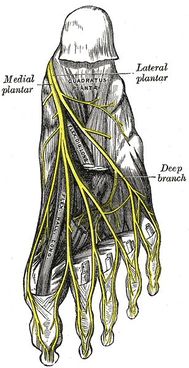
The lateral plantar nerve or the external plantar nerve ''(latin: nervus plantaris lateralis),'' it enters the sole of the [[Foot Anatomy|foot]] by passing deep to the proximal insertion of the [[Abductor Hallucis|abductor hallucis]] muscle. It continues across the sole anteriorly and laterally, between the flexor digitorum brevis and [[Quadratus Plantae|quadratus plantae]] muscles, innervating both of these muscles. Then, close to the 5th metatarsal head, it will divide into the deep and superficial branches<ref>Develi S.[https://pubmed.ncbi.nlm.nih.gov/29177688/ Trifurcation of the tibial nerve within the tarsal tunnel.]Surg Radiol Anat ''2018;''40.5: 529-532.</ref> | |||
=== Root === | |||
The lateral plantar nerve is a terminal branch of the [[Tibial Nerve|tibial nerve]]. | |||
=== Branches === | |||
* The superficial branch of the lateral plantar nerve splits into: | |||
** The lateral proper plantar digital nerve, which innervates the skin of the lateral aspects of the 5th toe, flexor digiti quinti brevis and interossei of the fourth web space<ref name=":2">De Maeseneer M, Madani H, Lenchik L, Kalume Brigido M, Shahabpour M, Marcelis S, De Mey J, Scafoglieri A. [https://d1wqtxts1xzle7.cloudfront.net/43175196/De_Maeseneer_RGPHS_2015-libre.pdf?1456704498=&response-content-disposition=inline%3B+filename%3DMUSCULOSKELETAL_IMAGING_Normal_Anatomy_a.pdf&Expires=1678731782&Signature=VpXMsEIkCad8DYoq2tIj0MB1P~A3kPBjhFx0pMUW~FdgAg-vpjBNn0bHCRwPS25aJSWEwok9YhzXrRsLSQn5y18hmRKJMGsQp5xXn1FSRQcxDbaF4JvDRqUXYzd~d6bEzDLFsxP4AGhNnX2PfqYnt3b~Lsbx9s95xYyu4dVatQffoCdqrK9hNglMuRMcANCLF4MjAiXN-OGjPKzrQz1KWUw4zcpy1Sc7dw24uclNYWmsHomyfFfJAyE0rinCXFVse4atgSpwy7bVOhRqGUec8BS0rguxKXjUPwIKck4RAkc5IhUakgVlk2N84-o6aHPQvAY0khIObyaoDKSpgaSp2w__&Key-Pair-Id=APKAJLOHF5GGSLRBV4ZA Normal anatomy and compression areas of nerves of the foot and ankle: US and MR imaging with anatomic correlation]. Radiographics. 2015 Sep;35(5):1469-82.</ref>. | |||
** The common plantar digital nerve may communicates with the 3rd common digital branch of the [[Medial Plantar Nerve|medial plantar nerve]] and forms the fourth common plantar digital nerve<ref name=":2" />. | |||
* The deep branch of the lateral plantar nerve innervates the interossei of the first three web spaces, the a[[Adductor Hallucis|dductor hallucis muscle]] and the 2nd, 3rd and 4th lumbrical muscles<ref name=":2" />. | |||
== Function == | |||
=== Motor === | |||
It is a motor nerve that innervates all the intrinsic muscles from the sole with the exception of abductor hallucis, flexor digitorum brevis, the flexor hallucis brevis, and the first lumbrical muscle innervated by the [[Medial Plantar Nerve|medial plantar nerve]]. | |||
[[File:Lateral plantar nerve distribution.png|thumb]] | |||
=== Sensory === | |||
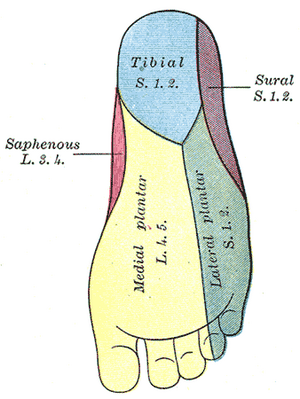
It also a sensory nerve that provides sensory information from the two anterior thirds of the lateral sole of the foot, as well as the plantar surfaces for the 5th and half of the 4th toe. | |||
== Clinical relevance == | |||
The entrapment of the lateral plantar nerve first branch (inferior calcaneal nerve), also known as Baxter's nerve, produces pain on the inside of the ankle and heel and can mimic [[Plantar Fasciitis|plantar fasciitis]] and [[Tarsal Tunnel Syndrome|tarsal tunnel syndrome]]<ref name=":1" />. Pain tends to increase with weight-bearing after prolonged rest or when rising in the morning. The pain may initially improve but then increases as the day progresses<ref name=":1" />. | |||
== Assessment == | |||
[[Baxter's Nerve Entrapment|Baxter's neuropathy]]:<ref name=":1">Thomas, Christensen JC, Kravitz SR, Mendicino RW, Schuberth JM, Vanore JV, et all. [https://pubmed.ncbi.nlm.nih.gov/20439021/ The diagnosis and treatment of heel pain: a clinical practice guideline-revision] . J Foot Ankle Surg 2010;49(3 Suppl):S1–S19. </ref><ref name=":0">Ferkel E, Davis WH, Ellington JK [https://pubmed.ncbi.nlm.nih.gov/26409596/ Entrapment neuropathies of the foot and ankle.] Clin sports med 2015; 34(4): 791-801.</ref> | |||
* Radiating pain may be present when the nerve is palpated. | |||
* There is maximal tenderness at the medial border of the heel where the entrapment occurs – usually around the origin and deep to the abductor hallucis. This may create radiating and/or burning pain laterally across the plantar foot. | |||
* There is a positive Phalen’s test (invert and plantarflex the foot passively). This compresses the nerve due to the narrowing of the porta pedis. | |||
* There may be weak abduction of the fifth toe. | |||
* There may be a positive Tinel’s sign. Pareasthesias may be reproduced with tapping over the nerve beneath the abductor hallucis muscle. | |||
* In chronic cases, patients may have diminished sensation in the lateral plantar foot. | |||
== Treatment == | |||
* Conservative treatment includes: <ref name=":0" /><ref>Davis PF, Severud E, Baxter DE. [https://pubmed.ncbi.nlm.nih.gov/7834059/ Painful heel syndrome: results of nonoperative treatment]. ''Foot Ankle Int''. 1994;15(10):531-535. </ref> | |||
# Taping and/or orthotics to control overpronation. | |||
# Stretching of the soleus and gastrocnemius muscles. | |||
# Soft tissue therapy to the plantar fascia and foot intrinsics. | |||
# Non-steroidal anti-inflammatory medication (NSAIDs). | |||
# Strengthening exercises for the foot intrinsics. | |||
* Non-conservative treatment:<ref>Lui TH. [https://link.springer.com/article/10.1007/s00402-007-0380-1#citeas Endoscopic decompression of the first branch of the lateral plantar nerve]. Arch Orthop Trauma Surg 2007;127(9): 859-861.</ref> | |||
# Endoscopic decompression of the first branch of the lateral plantar nerve | |||
== References == | |||
<references /> | |||
[[Category:Anatomy]] | |||
[[Category:Nerves]] | |||
__NEWSECTIONLINK__ | __NEWSECTIONLINK__ | ||
[[Category:Ankle - Anatomy]] | |||
[[Category:Nerves]] | |||
[[Category:Foot - Anatomy]] | |||
[[Category:Neuropathy]] | |||
Latest revision as of 20:02, 13 March 2023
Top Contributors - Elena Ferrero Vila, Leana Louw and Wendy Snyders
Description[edit | edit source]
The lateral plantar nerve or the external plantar nerve (latin: nervus plantaris lateralis), it enters the sole of the foot by passing deep to the proximal insertion of the abductor hallucis muscle. It continues across the sole anteriorly and laterally, between the flexor digitorum brevis and quadratus plantae muscles, innervating both of these muscles. Then, close to the 5th metatarsal head, it will divide into the deep and superficial branches[1]
Root[edit | edit source]
The lateral plantar nerve is a terminal branch of the tibial nerve.
Branches[edit | edit source]
- The superficial branch of the lateral plantar nerve splits into:
- The lateral proper plantar digital nerve, which innervates the skin of the lateral aspects of the 5th toe, flexor digiti quinti brevis and interossei of the fourth web space[2].
- The common plantar digital nerve may communicates with the 3rd common digital branch of the medial plantar nerve and forms the fourth common plantar digital nerve[2].
- The deep branch of the lateral plantar nerve innervates the interossei of the first three web spaces, the adductor hallucis muscle and the 2nd, 3rd and 4th lumbrical muscles[2].
Function[edit | edit source]
Motor[edit | edit source]
It is a motor nerve that innervates all the intrinsic muscles from the sole with the exception of abductor hallucis, flexor digitorum brevis, the flexor hallucis brevis, and the first lumbrical muscle innervated by the medial plantar nerve.
Sensory[edit | edit source]
It also a sensory nerve that provides sensory information from the two anterior thirds of the lateral sole of the foot, as well as the plantar surfaces for the 5th and half of the 4th toe.
Clinical relevance[edit | edit source]
The entrapment of the lateral plantar nerve first branch (inferior calcaneal nerve), also known as Baxter's nerve, produces pain on the inside of the ankle and heel and can mimic plantar fasciitis and tarsal tunnel syndrome[3]. Pain tends to increase with weight-bearing after prolonged rest or when rising in the morning. The pain may initially improve but then increases as the day progresses[3].
Assessment[edit | edit source]
- Radiating pain may be present when the nerve is palpated.
- There is maximal tenderness at the medial border of the heel where the entrapment occurs – usually around the origin and deep to the abductor hallucis. This may create radiating and/or burning pain laterally across the plantar foot.
- There is a positive Phalen’s test (invert and plantarflex the foot passively). This compresses the nerve due to the narrowing of the porta pedis.
- There may be weak abduction of the fifth toe.
- There may be a positive Tinel’s sign. Pareasthesias may be reproduced with tapping over the nerve beneath the abductor hallucis muscle.
- In chronic cases, patients may have diminished sensation in the lateral plantar foot.
Treatment[edit | edit source]
- Taping and/or orthotics to control overpronation.
- Stretching of the soleus and gastrocnemius muscles.
- Soft tissue therapy to the plantar fascia and foot intrinsics.
- Non-steroidal anti-inflammatory medication (NSAIDs).
- Strengthening exercises for the foot intrinsics.
- Non-conservative treatment:[6]
- Endoscopic decompression of the first branch of the lateral plantar nerve
References[edit | edit source]
- ↑ Develi S.Trifurcation of the tibial nerve within the tarsal tunnel.Surg Radiol Anat 2018;40.5: 529-532.
- ↑ 2.0 2.1 2.2 De Maeseneer M, Madani H, Lenchik L, Kalume Brigido M, Shahabpour M, Marcelis S, De Mey J, Scafoglieri A. Normal anatomy and compression areas of nerves of the foot and ankle: US and MR imaging with anatomic correlation. Radiographics. 2015 Sep;35(5):1469-82.
- ↑ 3.0 3.1 3.2 Thomas, Christensen JC, Kravitz SR, Mendicino RW, Schuberth JM, Vanore JV, et all. The diagnosis and treatment of heel pain: a clinical practice guideline-revision . J Foot Ankle Surg 2010;49(3 Suppl):S1–S19.
- ↑ 4.0 4.1 Ferkel E, Davis WH, Ellington JK Entrapment neuropathies of the foot and ankle. Clin sports med 2015; 34(4): 791-801.
- ↑ Davis PF, Severud E, Baxter DE. Painful heel syndrome: results of nonoperative treatment. Foot Ankle Int. 1994;15(10):531-535.
- ↑ Lui TH. Endoscopic decompression of the first branch of the lateral plantar nerve. Arch Orthop Trauma Surg 2007;127(9): 859-861.


