Supinator: Difference between revisions
(replace wrong ampersand with single one) |
Kim Jackson (talk | contribs) m (Text replacement - "biceps brachii" to "Biceps Brachii") |
||
| (12 intermediate revisions by 3 users not shown) | |||
| Line 2: | Line 2: | ||
'''Original Editor '''- [[User:Kate Sampson|Kate Sampson]], | '''Original Editor '''- [[User:Kate Sampson|Kate Sampson]], | ||
'''Top Contributors''' - {{Special:Contributors/{{FULLPAGENAME}}}}; | '''Top Contributors''' - {{Special:Contributors/{{FULLPAGENAME}}}}; | ||
</div> | |||
= Anatomy | == Description == | ||
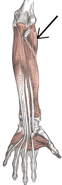
[[File:Supinator muscle.png|thumb|[https://commons.wikimedia.org/wiki/File:Musculussupinator.png Supinator muscle]]] | |||
The supinator muscle is located in the posterior compartment of the forearm. As its name suggests, it is the prime supinator of the forearm.<ref name=":0">Gray H. ''[https://www.bartleby.com/107/125.html Anatomy of the Human Body.]'' Twentieth edition. Philadelphia: Lea & Febiger; 918 Available from: https://www.bartleby.com/107/125.html [Accessed 23 May 2019]</ref><br> | |||
== | == Structure == | ||
The supinator is a broadly-shaped muscle in the superior and posterior compartment of the forearm, It curves around the upper third of the radius and consists of two layers of fibres. In between these layers lies the deep branch of the [[radial nerve]].<ref name=":0" /> | |||
== Origin == | |||
The supinator muscle has a broad origin from the [[ulna]] and [[humerus]]. The two layers of fibres originate in common from the supinator crest of the [[ulna]], the lateral epicondyle of the [[humerus]], the radial collateral ligament and the annular radial ligament. The superficial layer of fibres has a tendinous origin and surround the upper part of the radius. The deeper layer of fibres encircles the neck of the [[radius]] above the radial tuberosity.<ref name="Martini" /> | |||
== Insertion == | |||
Proximal third of the [[radius]] on the anterolateral and posterior surface, distal to the radial tuberosity. <ref name="Martini">Martini FH. ''Fundamentals of Anatomy and Physiology''. Seventh Edition. Pearson Education Inc; 2006.</ref> | |||
== | == Innervation == | ||
The supinator muscle is innervated by the deep branch of the [[radial nerve]], that becomes the posterior interosseous nerve upon exiting the supinator muscle (C5, C6) <ref name="Palastanga">Palastanga N, Field D, Soames R. ''Anatomy and human movement. Structure and function''. 5th ed. London: Butterworth Heinemann Elsevier; 2006</ref> | |||
== | == Function == | ||
The main function of the supinator is to supinate the forearm. This can be done with the elbow in any position of flexion or extension. Supinator works with [[Biceps Brachii]] if powerful supination is required. However, [[Biceps Brachii]] is unable to supinate when the elbow is extended.<ref name="Palastanga" /> | |||
== Clinical Relevance == | |||
The radial nerve divides into deep and sensory superficial branches just proximal to the supinator muscle. This can lead to entrapment and compression of the deep branch and could potentially result in selective paralysis of the muscles served by this nerve. Many possible causes are known for this nerve syndrome (known as supinator entrapment syndrome) including compression by various soft-tissued masses surrounding the nerve, and stress caused by repetitive supination and pronation<br> | |||
= | == Assessment == | ||
Place the patient's arm and elbow in extension with the forearm in mid-position. Actively resist supination and palpate along the posterior part of the proximal third of the radius. <ref name="Palastanga" /> | |||
== Resources == | |||
{{#ev:youtube|4FdX75utp0Y}} | |||
== | == References == | ||
<references /> | <references /> | ||
[[Category:Muscles]] | [[Category:Muscles]] | ||
[[Category:Anatomy]] | |||
[[Category:Anatomy Project]] | |||
[[Category:Elbow]] | |||
[[Category:Elbow - Anatomy]] | |||
[[Category:Elbow - Muscles]] | |||
Latest revision as of 14:57, 10 January 2022
Original Editor - Kate Sampson,
Top Contributors - Kate Sampson, Kim Jackson, Nina Myburg, 127.0.0.1 and Joao Costa;
Description[edit | edit source]
The supinator muscle is located in the posterior compartment of the forearm. As its name suggests, it is the prime supinator of the forearm.[1]
Structure[edit | edit source]
The supinator is a broadly-shaped muscle in the superior and posterior compartment of the forearm, It curves around the upper third of the radius and consists of two layers of fibres. In between these layers lies the deep branch of the radial nerve.[1]
Origin[edit | edit source]
The supinator muscle has a broad origin from the ulna and humerus. The two layers of fibres originate in common from the supinator crest of the ulna, the lateral epicondyle of the humerus, the radial collateral ligament and the annular radial ligament. The superficial layer of fibres has a tendinous origin and surround the upper part of the radius. The deeper layer of fibres encircles the neck of the radius above the radial tuberosity.[2]
Insertion[edit | edit source]
Proximal third of the radius on the anterolateral and posterior surface, distal to the radial tuberosity. [2]
Innervation[edit | edit source]
The supinator muscle is innervated by the deep branch of the radial nerve, that becomes the posterior interosseous nerve upon exiting the supinator muscle (C5, C6) [3]
Function[edit | edit source]
The main function of the supinator is to supinate the forearm. This can be done with the elbow in any position of flexion or extension. Supinator works with Biceps Brachii if powerful supination is required. However, Biceps Brachii is unable to supinate when the elbow is extended.[3]
Clinical Relevance[edit | edit source]
The radial nerve divides into deep and sensory superficial branches just proximal to the supinator muscle. This can lead to entrapment and compression of the deep branch and could potentially result in selective paralysis of the muscles served by this nerve. Many possible causes are known for this nerve syndrome (known as supinator entrapment syndrome) including compression by various soft-tissued masses surrounding the nerve, and stress caused by repetitive supination and pronation
Assessment[edit | edit source]
Place the patient's arm and elbow in extension with the forearm in mid-position. Actively resist supination and palpate along the posterior part of the proximal third of the radius. [3]
Resources[edit | edit source]
References[edit | edit source]
- ↑ 1.0 1.1 Gray H. Anatomy of the Human Body. Twentieth edition. Philadelphia: Lea & Febiger; 918 Available from: https://www.bartleby.com/107/125.html [Accessed 23 May 2019]
- ↑ 2.0 2.1 Martini FH. Fundamentals of Anatomy and Physiology. Seventh Edition. Pearson Education Inc; 2006.
- ↑ 3.0 3.1 3.2 Palastanga N, Field D, Soames R. Anatomy and human movement. Structure and function. 5th ed. London: Butterworth Heinemann Elsevier; 2006


