Upper Extremity Myofascial Chains: Difference between revisions
No edit summary |
No edit summary |
||
| Line 13: | Line 13: | ||
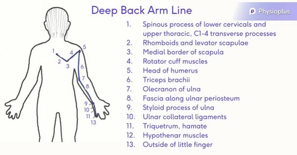
# Deep back arm line (figure 4) | # Deep back arm line (figure 4) | ||
<gallery widths=" | <gallery widths="350px" heights="250px"> | ||
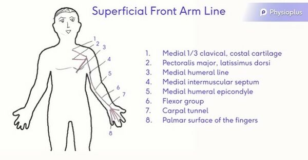
File: Superficial Front Arm Line (1).jpg|600x600px|alt=|thumb|Figure 1. Superficial front arm line. | File: Superficial Front Arm Line (1).jpg|600x600px|alt=|thumb|Figure 1. Superficial front arm line. | ||
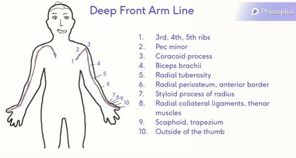
File: Deep Front Arm Line (2).jpg|600x600px|alt=|thumb|Figure 2. Deep front arm line. </gallery> | File: Deep Front Arm Line (2).jpg|600x600px|alt=|thumb|Figure 2. Deep front arm line. </gallery> | ||
Revision as of 12:43, 12 September 2021
Top Contributors - Jess Bell, Carin Hunter, Kim Jackson, Lucinda hampton and Robin Tacchetti
Introduction[edit | edit source]
A myofascial chain is a line of connective tissue that runs throughout the body. There is a posterior (back) line, an anterior (front) line, a spiral line, and a lateral line. These lines help the body to move as a unit.[1]
Myofascial chains are important for functional movement, coordination and stability.[2] They can, however, also cause pain within the body and structural weakness.[3] By understanding myofascial chains, you can better understand injuries and movement limitations.[4]
In the upper limb there are four myofascial chains[5]:
- Superficial front arm line (figure 1)
- Deep front arm line (figure 2)
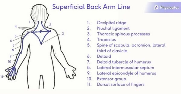
- Superficial back arm line (figure 3)
- Deep back arm line (figure 4)
Stabilisation Tracts[edit | edit source]
1. Back arm line[edit | edit source]
- Latissimus dorsi
- Thoracolumbar fascia
- Sacral fascia contralateral to thoracolumbar fascia
- Gluteus max contralateral to thoracolumbar fascia
- Vastus lateralis
2. Front arm line[edit | edit source]
- Pectoralis major
- External oblique
- Adductor longus contralateral to external oblique
- Gracilis contralateral to external oblique
- Pes anserine contralateral to external oblique
- Tibial periosteum contralateral to external oblique
References[edit | edit source]
- ↑ Bordoni B, Myers T. A review of the theoretical fascial models: biotensegrity, fascintegrity, and myofascial chains. Cureus. 2020 Feb;12(2).
- ↑ Kazakos D, Liapis A, Mylonas K, Angelopoulos P, Koubetsos A, Tsepis E, Fousekis K. Treatment of scalene muscles with the Ergon technique can lead to greater improvement in hip abduction range of motion than local hip adductor treatment: a study on deep front line connectivity. Journal of Physical Therapy Science. 2020;32(11):706-9.
- ↑ Dischiavi SL, Wright AA, Hegedus EJ, Bleakley CM. Biotensegrity and myofascial chains: A global approach to an integrated kinetic chain. Medical hypotheses. 2018 Jan 1;110:90-6.
- ↑ Burk C, Perry J, Lis S, Dischiavi S, Bleakley C. Can myofascial interventions have a remote effect on ROM? A systematic review and meta-analysis. Journal of sport rehabilitation. 2019 Oct 18;29(5):650-6.
- ↑ Wilke J, Krause F. Myofascial chains of the upper limb: a systematic review of anatomical studies. Clinical Anatomy. 2019 Oct;32(7):934-40.


