Lymphatic System: Difference between revisions
No edit summary |
No edit summary |
||
| Line 47: | Line 47: | ||
* Tonsils - Oval-shaped masses of glandular tissue located on both sides at the back of the throat. Tonsils act like filters to trap bacteria and viruses.<ref>Gale Encyclopedia of Medicine. Copyright 2008 The Gale Group, Inc. All rights reserved.Available from:https://medical-dictionary.thefreedictionary.com/Tonsils (last accessed 27.7.2020)</ref> | * Tonsils - Oval-shaped masses of glandular tissue located on both sides at the back of the throat. Tonsils act like filters to trap bacteria and viruses.<ref>Gale Encyclopedia of Medicine. Copyright 2008 The Gale Group, Inc. All rights reserved.Available from:https://medical-dictionary.thefreedictionary.com/Tonsils (last accessed 27.7.2020)</ref> | ||
3. Other lymphoid tissue (or diffuse lymphoid regions not encapsulated)<ref>Preserve articles [https://www.preservearticles.com/biology/what-are-secondary-lymphoid-organs/25163 What are Secondary Lymphoid Organs?] Available from:https://www.preservearticles.com/biology/what-are-secondary-lymphoid-organs/25163 (last accessed 27.7.2020)</ref> | 3. Other lymphoid tissue (or diffuse lymphoid regions not encapsulated)<ref>Preserve articles [https://www.preservearticles.com/biology/what-are-secondary-lymphoid-organs/25163 What are Secondary Lymphoid Organs?] Available from:https://www.preservearticles.com/biology/what-are-secondary-lymphoid-organs/25163 (last accessed 27.7.2020)</ref> | ||
* Mucosa-associated lymphoid tissue (MALT) is an umbrella term for extranodal aggregates of lymphoid tissue in the bronchus (BALT), gut (GALT) and skin (SALT), as well as breast and uterine cervix. MALT is the arm of the immune defence in closest contact with exogenous antigens, thus differing from the compartmentalised peripheral somatic lymphoid tissues, which include the lymph nodes, thymus and spleen. It constitutes about 50 percent of the lymphoid tissue in the human body<ref>careers 300 [https://learn.careers360.com/medical/question-i-have-a-doubt-kindly-clarify-malt-constitutes-about_________________-percent-of-thelymphoid-tissue-in-human-body/ MALT] Available from:https://learn.careers360.com/medical/question-i-have-a-doubt-kindly-clarify-malt-constitutes-about_________________-percent-of-thelymphoid-tissue-in-human-body/ (last accessed 27.7.2020)</ref><ref name=":3" /><ref>medical dictionary MALT Available from:https://medical-dictionary.thefreedictionary.com/mucosa-associated+lymphoid+tissue (last accessed 27.7.2020)</ref> | *[[File:2210 Mucosa Associated Lymphoid Tissue (MALT) Nodule.jpg|right|frameless|450x450px]]Mucosa-associated lymphoid tissue (MALT) is an umbrella term for extranodal aggregates of lymphoid tissue in the bronchus (BALT), gut (GALT) and skin (SALT), as well as breast and uterine cervix. MALT is the arm of the immune defence in closest contact with exogenous antigens, thus differing from the compartmentalised peripheral somatic lymphoid tissues, which include the lymph nodes, thymus and spleen. It constitutes about 50 percent of the lymphoid tissue in the human body<ref>careers 300 [https://learn.careers360.com/medical/question-i-have-a-doubt-kindly-clarify-malt-constitutes-about_________________-percent-of-thelymphoid-tissue-in-human-body/ MALT] Available from:https://learn.careers360.com/medical/question-i-have-a-doubt-kindly-clarify-malt-constitutes-about_________________-percent-of-thelymphoid-tissue-in-human-body/ (last accessed 27.7.2020)</ref><ref name=":3" /><ref>medical dictionary MALT Available from:https://medical-dictionary.thefreedictionary.com/mucosa-associated+lymphoid+tissue (last accessed 27.7.2020)</ref> | ||
At R - Mucosa Associated Lymphoid Tissue (MALT) Nodule. Peyer's patches (any of numerous large oval patches of closely aggregated nodules of lymphoid tissue in the walls of the small intestine especially in the ileum that partially or entirely disappear in advanced life and in typhoid fever become the seat of ulcers which may perforate the intestines<ref>Merrium Webster [https://www.merriam-webster.com/medical/Peyer's%20patch Peyers Patche]s Available from:https://www.merriam-webster.com/medical/Peyer's%20patch (last accessed 27.7.2020)</ref>) and mucosa illustrated. | |||
== Diseases of the Lymphatic System == | == Diseases of the Lymphatic System == | ||
Revision as of 07:36, 27 July 2020
This article is currently under review and may not be up to date. Please come back soon to see the finished work! (27/07/2020)
Introduction[edit | edit source]

The lymphatic system is an important and often underappreciated component of the circulatory, immune, and metabolic systems. Lymphatic system is considered as a part of both the circulatory and immune systems. The functions of the lymphatic system complement the bloodstream functions, as it regulates the balance of fluids in the body and filters the pathogens from the blood[1].
It is composed of
- Lymphatic fluid - protein rich fluid that flows through the lymphatic system and surrounds all tissues [2]. It carries about 75-100 grams of protein circulating in the body per day. Lymph is composed of white blood cells, triglycerides, bacteria, cell debris, water, and protein. It has a composition comparable to blood plasma.
- Lymphatic vessels - reabsorb interstitial fluid from the periphery to return it to the intravascular space, which prevents fluid build up in peripheral tissues
- Lymphatic cells - include macrophages, dendritic cells, lymphocytes
- Lymphatic organs - eg spleen and thymus.
There are three primary functions of the lymphatic system:
- Maintenance of fluid balance
- Facilitation of the absorption of dietary fats from the gastrointestinal tract to the bloodstream for metabolism or storage
- Enhancement and facilitation of the immune system[3].
Problems that lymph may cause are primarily because there is no pump in the system[4].
Lymphatic Transport System[edit | edit source]

5 components: capillaries, collecting vessels, lymph nodes, trunks, and ducts.
- The initial point of entry into the lymphatic system is through the capillaries - a single layer of partially overlapping endothelial cells creating a valve. These overlapping junctions form a "button-like" opening, which allows fluid into the capillary when the pressure outside of the vessel is greater than the pressure inside of the vessel. The capillaries subsequently feed fluid into the collecting vessels.
- Collecting vessels are composed of an endothelial layer with many tight junctions forming a "zipper-like" structure. These collecting vessels also have intraluminal valves as well as pericytes. The pericytes contain alpha-smooth muscle actin, which functions to contract the vessel and pump the fluid further through the system. The valves prevent the backflow of the lymphatic fluid and ensure the unidirectional flow of fluid.
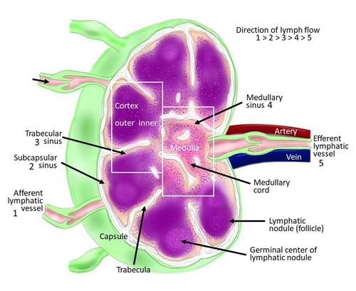
- 3. Lymph nodes (see image R) - are the next point in the system. They possess a medulla, paracortex, and cortex which in combination, form the house of lymphocytes, antigen-presenting cells, and macrophages. There are approximately 450 lymph nodes in the human body. After visiting the lymph nodes, the lymphatic fluid flows into efferent lymphatic collecting vessels
- Lymphatic trunks are the next vessels
- Lymphatic ducts - Either of the two terminal lymph vessels that convey lymph to the bloodstream: right and left.

Left Duct (or thoracic duct) collects most of the lymph in the body other than from the right thorax, arm, head, and neck which are drained by the right lymphatic duct. It is 38–45 cm in length
Right Duct drains (1.25 cm in length) lymph fluid from:
- the upper right section of the trunk, (right thoracic cavity, via the right bronchomediastinal trunk),
- the right arm (via the right subclavian trunk),
- and right side of the head and neck (via the right jugular trunk)[5],
The lymphatic ducts allow the entry of the lymphatic fluid into the venous system bilaterally via the opening found at the intersection of the subclavian and internal jugular vein[3].
Lymphatic Organs and Tissues[edit | edit source]
- Primary lymphoid organs
The primary lymphoid organs generate lymphocytes from immature progenitor cells. The thymus and the bone marrow constitute the primary lymphoid organs involved in the production and early clonal selection of lymphocyte tissues.
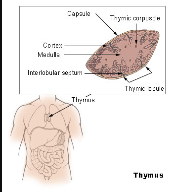
- Thymus - a ductless gland lying in the upper mediastinum beneath the sternum; reaches its maximum development during puberty and continues to play an immunologic role throughout life - though its function declines with age. During the last stages of fetal life and the early neonatal period, the reticular structure of the thymus entraps immature stem cells arising from the bone marrow and circulating in the blood. The thymus preprocesses these cells, causing them to become sensitized and capable of maturing into a specific differentiated type of lymphocyte. After sensitization by the thymus, the cells reenter the blood and are transported to developing lymphoid tissue, where they seed the cells that eventually become T lymphocytes (essential to the development of cell-mediated immunity).[6].
- Bone marrow - soft, organic, spongelike material in the cavities of bones. It is a network of blood vessels and special connective tissue fibers that hold together a composite of fat and blood-producing cells. Chief function is to manufacture erythrocytes, leukocytes, and platelets. These blood cells normally do not enter the bloodstream until they are fully developed, so that the marrow contains cells in all stages of growth. If the body's demand for leukocytes is increased because of infection, the marrow responds immediately by stepping up production.[7]

2. Secondary lymphoid organs
The secondary lymphoid organs may be encapsulated to have a specific shape or may be in the form of diffused tissues. Spleen, lymph nodes, adenoids, tonsils are the encapsulated secondary lymphoid organs. They maintain mature naive lymphocytes and initiate an adaptive immune response. The peripheral lymphoid organs are the sites of lymphocyte activation by antigens. Activation leads to clonal expansion and affinity maturation. Mature lymphocytes recirculate between the blood and the peripheral lymphoid organs until they encounter their specific antigen[5].
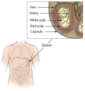
- Spleen - An organ that is located in the upper-left part of the abdomen, not far from the stomach, that produces lymphocytes, which are important elements in the immune system. The spleen is the largest lymphatic organ in the body. The spleen also filters blood, serves as a major reservoir for blood, and destroys blood cells that are aged (or abnormal, as in the case of sickle cells)[8]
- Lymph node any of the accumulations of lymphoid tissue organized as definite lymphoid organs along the course of lymphatic vessels; they consist of an outer cortical and an inner medullary part. Lymph nodes are the main source of lymphocytes of the peripheral blood and, as part of the reticuloendothelial system, serve as a defense mechanism by removing noxious agents such as bacteria and toxins, and probably play a role in antibody formation[9].
- Adenoids - A normal collection of unencapsulated lymphoid tissue in the nasopharynx. Also called pharyngeal tonsils.[10]
- Tonsils - Oval-shaped masses of glandular tissue located on both sides at the back of the throat. Tonsils act like filters to trap bacteria and viruses.[11]

3. Other lymphoid tissue (or diffuse lymphoid regions not encapsulated)[12]
- Mucosa-associated lymphoid tissue (MALT) is an umbrella term for extranodal aggregates of lymphoid tissue in the bronchus (BALT), gut (GALT) and skin (SALT), as well as breast and uterine cervix. MALT is the arm of the immune defence in closest contact with exogenous antigens, thus differing from the compartmentalised peripheral somatic lymphoid tissues, which include the lymph nodes, thymus and spleen. It constitutes about 50 percent of the lymphoid tissue in the human body[13][5][14]

_Nodule.jpg)
At R - Mucosa Associated Lymphoid Tissue (MALT) Nodule. Peyer's patches (any of numerous large oval patches of closely aggregated nodules of lymphoid tissue in the walls of the small intestine especially in the ileum that partially or entirely disappear in advanced life and in typhoid fever become the seat of ulcers which may perforate the intestines[15]) and mucosa illustrated.
Diseases of the Lymphatic System[edit | edit source]

The lymphatic system is also prone to diseases like the venous and arterial circulation. Eg
- Lymphedema. When this occurs, the lymphatic system is unable to drain lymphatic fluid which results in accumulation of the fluid causing swelling of the extremity. Lymphedema is classified as primary or secondary.
- Primary lymphedema is an inherited disorder where the lymphatics may be missing or abnormally developed (eg. aplasia, hypoplasia, hyperplasia). This condition usually presents at birth or sometimes may present later in life.
- Secondary lymphedema is an acquired disorder that has a known cause such as cancer, infection, trauma or following a surgical procedure.
- Lymphomas are malignancies that arise from the cells of the lymphatic system. There is usually malignant transformation of specific lymphocytes in the lymphatics or lymph nodes that are present in the gastrointestinal tract, neck, axilla or groin. Symptoms of lymphoma may include night sweats, fever, fatigue, itching and weight loss.
- Cancers of a variety of organs may commonly spread to involve regional lymph nodes.
- Lymphadenitis occurs when the lymph nodes become inflamed. The cause is usually an adjacent bacterial infection. The lymph nodes usually enlarge and become tender.
- Filariasis is a very common disorder caused by a parasite in Africa. The parasite rapidly divided and obstructs the lymph nodes in the groin, making it difficult for the lymphatics to drain the extremity. This often results in huge extremities and marked disability.[16]
Physiotherapy[edit | edit source]
Physiotherapy has a crucial part in helping to resolve problems with the lymphatic system such as using manual lymphatic drainage and exercise which are components of Complete Decongestive Therapy (CDT) [17],[4], [18] [19]. Exercise induces transport of lymph. Movement of muscles and arterial pulses cause the transport of lymph through the lymphatic system. Being active therefore has a positive effect of lymphatic system and the movement of lymph through our body. Some activities that are recommended are swimming, brisk walking, tennis, jumping, gymnastic, deep breathing, etc [20] [21]
Manual lymph drainage has shown to be of benefit in examples below
- Hind foot operations:the application of lymph drainage techniques after hindfoot (the posterior part of the human foot that contains the calcaneus, talus, navicular, and cuboid bones) operations, in combination with standard physiotherapy exercises, achieves greater limb volume reduction than exercise alone. [22]
- Breast cancer related lymph edema:[23] [24]
- Sports Medicine and Rehabilitation.[25]
Key Points[edit | edit source]
- Lymph nodes function to monitor the composition of lymphatic fluid/blood, engulf any pathogens, augment an immune response, and eradicate infection. They are like filtering stations for the lymph fluid.
- The thymus serves to mature and develop T cells in response to an inflammatory process, immune response or malignancy.
- The absorption and transport of fats and fat-soluble vitamins from the GI system also requires lymphatics.
- The lymphatic fluid is eventually emptied at the junction of the left subclavian vein and left internal jugular veins.
- Lymphatic fluid is derived from plasma. It leaks out of the capillary walls because of pressure exerted by the heart or osmotic pressure at the cellular level.
- In the GI tract, the lymphatic fluid has a milk-like appearance that is chiefly due to the presence of cholesterol, glycerol, fatty acids and other fat products. The vessels that transport the lymphatic fluid from the GI tract are known as lacteals.
- Lymphatic capillaries are very thin vessels which are blind-ended tubes. The lymphatic capillaries tend to form a large network of tubes that are known as lymphatic vessels.
- The key feature of lymphatic vessels is that they have thin endothelial walls and have an overlapping arrangement. This morphology allows for any fluid from the tissues to enter the cells. [16]
References[edit | edit source]
- ↑ Ken Hub Lymphatic system Available from:https://www.kenhub.com/en/library/anatomy/lymphatic-system (last accessed 27.7.2020)
- ↑ Foeldi M. Anatomy of the Lymphatic System. In Foeldi M, Foeldi Textbook of Lymphology. Munich: Urban and Fischer; 2006: Chapter 1.
- ↑ 3.0 3.1 Ozdowski L, Gupta V. Physiology, Lymphatic System. InStatPearls [Internet] 2020 May 21. StatPearls Publishing.Available from:https://www.statpearls.com/kb/viewarticle/24563 (last accessed 27.7.2020)
- ↑ 4.0 4.1 L.Felts. Detox your lymph: 10 holistic treatments for your lymphatic system. Available from: https://thechalkboardmag.com/detox-your-lymph-10-holistic-treatments-for-lymphatic-system (accessed 13/02/2019)
- ↑ 5.0 5.1 5.2 Wikipedia Lymphatics Available from:https://en.wikipedia.org/wiki/Lymphatic_system (last accessed 27.7.2020)
- ↑ Medical dictionary Thymus Available from:https://medical-dictionary.thefreedictionary.com/thymus (last accessed 27.7.2020)
- ↑ Med dictionary Bone marrow Available from:https://medical-dictionary.thefreedictionary.com/bone+marrow (last accessed 27.7.2020)
- ↑ medicine net Spleen Available from:https://www.medicinenet.com/script/main/art.asp?articlekey=5531 (last accessed 27.7.2020)
- ↑ https://medical-dictionary.thefreedictionary.com/lymph+node
- ↑ Medical Dictionary for the Health Professions and Nursing © Farlex 2012 Available from:https://medical-dictionary.thefreedictionary.com/adenoids (last accessed 27.7.2020)
- ↑ Gale Encyclopedia of Medicine. Copyright 2008 The Gale Group, Inc. All rights reserved.Available from:https://medical-dictionary.thefreedictionary.com/Tonsils (last accessed 27.7.2020)
- ↑ Preserve articles What are Secondary Lymphoid Organs? Available from:https://www.preservearticles.com/biology/what-are-secondary-lymphoid-organs/25163 (last accessed 27.7.2020)
- ↑ careers 300 MALT Available from:https://learn.careers360.com/medical/question-i-have-a-doubt-kindly-clarify-malt-constitutes-about_________________-percent-of-thelymphoid-tissue-in-human-body/ (last accessed 27.7.2020)
- ↑ medical dictionary MALT Available from:https://medical-dictionary.thefreedictionary.com/mucosa-associated+lymphoid+tissue (last accessed 27.7.2020)
- ↑ Merrium Webster Peyers Patches Available from:https://www.merriam-webster.com/medical/Peyer's%20patch (last accessed 27.7.2020)
- ↑ 16.0 16.1 Null M, Agarwal M. Anatomy, Lymphatic System. InStatPearls [Internet] 2019 Jun 22. StatPearls Publishing. Available from:https://www.ncbi.nlm.nih.gov/books/NBK513247/ (last accessed15.2.2020)
- ↑ A. Weil. Lymphatic Massage Therapy. Available from: https://www.drweil.com/health-wellness/balanced-living/wellness-therapies/lymphatic-massage-therapy/ (accessed 12/02/2019)
- ↑ Boon C.C. Manual Lymphatic Drainage. Available from: https://www.physio-pedia.com/manual_lymph_drainage (accessed 11/02/2019)
- ↑ Dіdem K, Ufuk YS, Serdar S, Zümre A. The comparison of two different physiotherapy methods in treatment of lymphedema after breast surgery. Breast cancer research and treatment. 2005 Sep 1;93(1):49-54.
- ↑ L. Vandermeeren, G. Van Damme, S. Stein, N. Devoogdt, B. Clerinks, P. Vlecken, L. Martens, K. Tollenaere, N. Beauloye. Report Patient Day 29 october 2017. Available from: https://www.belymph.org/web/en/belymph/patient-day (accessed 16/02/2019)
- ↑ S. Ding. 16 warning sings that you need to drain your lymphatic fluids. Available from: https://juicing-for-health.com/how-to-drain-your-lymphatic-fluids (accessed 12/02/2019)
- ↑ Kessler T, de Bruin E, Brunner F, Vienne P, Kissling R. Effect of manual lymph drainage after hindfoot operations. Physiotherapy Research International. 2003 Jun;8(2):101-10.
- ↑ Atalay OT, Özkir A, Çalik BB, Baskan E, Taşkin H. Effects of phase I complex decongestive physiotherapy on physical functions and depression levels in breast cancer related lymph edema. Journal of physical therapy science. 2015;27(3):865-70.
- ↑ Lacomba MT, Sánchez MJ, Goñi ÁZ, Merino DP, del Moral OM, Téllez EC, Mogollón EM. Effectiveness of early physiotherapy to prevent lymphoedema after surgery for breast cancer: randomised, single blinded, clinical trial. Bmj. 2010 Jan 13;340:b5396.
- ↑ Vairo GL, Miller SJ, Rier NC, Uckley WI. Systematic review of efficacy for manual lymphatic drainage techniques in sports medicine and rehabilitation: an evidence-based practice approach. Journal of Manual & Manipulative Therapy. 2009 Jul 1;17(3):80E-9E.

