Navicular stress syndrome: Difference between revisions
Sazia Queyam (talk | contribs) m (Added images) |
Kim Jackson (talk | contribs) (Added under review banner) |
||
| (14 intermediate revisions by 2 users not shown) | |||
| Line 1: | Line 1: | ||
<div class="noeditbox"> | |||
This article is currently under review and may not be up to date. Please come back soon to see the finished work! ({{REVISIONDAY}}/{{REVISIONMONTH}}/{{REVISIONYEAR}}) | |||
</div> | |||
< | == Introduction == | ||
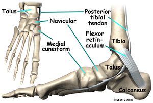
The Navicular is an intermediate tarsal bone on the medial side of the foot<ref>D.Richard, V.Wayne, M. Adam, Gray’s Anatomy for Students. Spain: Elsevier Publishers, 2005</ref>. Its name '''(''os naviculare pedis; scaphoid bone'')''' derives from the human bone's resemblance to a small boat. It articulates with ''four'' bones: the talus and the three cuneiforms; occasionally with a fifth, the cuboid. | |||
While rare in the general population, stress fractures of the tarsal navicular bone are frequently incurred by professional athletes.<ref>Shakked RJ, Walters EE, O’Malley MJ. [https://www.ncbi.nlm.nih.gov/pmc/articles/PMC5344863/ Tarsal navicular stress fractures.] Current reviews in musculoskeletal medicine. 2017 Mar 1;10(1):122-30.</ref> | |||
[[File:Foot accessory navicular CLINICAL ANATOMY 1 anat01.jpg|Navicular Bone.|center|frameless]] | |||
== Clinically Relevant Anatomy == | |||
=== | The navicular unique anatomic location subjects it to medial and lateral compression forces from the first and second meta-tarso-cuneiform joints, respectively<ref name=":0">Gross CE, Nunley JA. Navicular stress fractures. Foot & ankle international. 2015 Sep;36(9):1117-22.</ref>. While the medial forces are shared with the talar head, the lateral forces are borne by the navicular alone. As a result of this unequal distribution of forces, maximum sheer stress is concentrated at the central third of the bone<ref>Hossain M, Clutton J, Ridgewell M, Lyons K, Perera A. Stress fractures of the foot. Clinics in sports medicine. 2015 Oct 1;34(4):769-90.</ref><ref>McInnis KC, Ramey LN. High-risk stress fractures: diagnosis and management. PM&R. 2016 Mar 1;8(3):S113-24.</ref>. Moreover, contraction of the tibialis posterior tendon, which inserts on the navicular’s medial tuberosity elevates the medial stress experienced by the navicular<ref name=":0" />. With repeated compression of the navicular such as during running, the majority of forces are concentrated at the center of the bone, causing the navicular to be repeatedly “bent” at the avascular zone<ref>Mann G, Hetsroni I, Constantini N, Dolev E, Palmanovich E, Finsterbush A, Keltz E, Mei-Dan O, Eshed I, Marom N, Nyska M. Navicular stress fractures of the foot. Sports Injuries: Prevention, Diagnosis, Treatment and Rehabilitation. 2015:1-2.</ref><ref>Bennell K, Matheson G, Meeuwisse W, Brukner P. Risk factors for stress fractures. Sports medicine. 1999 Aug 1;28(2):91-122.</ref>. | ||
== References == | |||
<references /> | <references /> | ||
__INDEX__ | __INDEX__ | ||
[[Category:Sports Medicine]] | |||
[[Category:Sports Injuries]] | |||
Latest revision as of 03:11, 3 April 2020
This article is currently under review and may not be up to date. Please come back soon to see the finished work! (3/04/2020)
Introduction[edit | edit source]
The Navicular is an intermediate tarsal bone on the medial side of the foot[1]. Its name (os naviculare pedis; scaphoid bone) derives from the human bone's resemblance to a small boat. It articulates with four bones: the talus and the three cuneiforms; occasionally with a fifth, the cuboid.
While rare in the general population, stress fractures of the tarsal navicular bone are frequently incurred by professional athletes.[2]
Clinically Relevant Anatomy[edit | edit source]
The navicular unique anatomic location subjects it to medial and lateral compression forces from the first and second meta-tarso-cuneiform joints, respectively[3]. While the medial forces are shared with the talar head, the lateral forces are borne by the navicular alone. As a result of this unequal distribution of forces, maximum sheer stress is concentrated at the central third of the bone[4][5]. Moreover, contraction of the tibialis posterior tendon, which inserts on the navicular’s medial tuberosity elevates the medial stress experienced by the navicular[3]. With repeated compression of the navicular such as during running, the majority of forces are concentrated at the center of the bone, causing the navicular to be repeatedly “bent” at the avascular zone[6][7].
References[edit | edit source]
- ↑ D.Richard, V.Wayne, M. Adam, Gray’s Anatomy for Students. Spain: Elsevier Publishers, 2005
- ↑ Shakked RJ, Walters EE, O’Malley MJ. Tarsal navicular stress fractures. Current reviews in musculoskeletal medicine. 2017 Mar 1;10(1):122-30.
- ↑ 3.0 3.1 Gross CE, Nunley JA. Navicular stress fractures. Foot & ankle international. 2015 Sep;36(9):1117-22.
- ↑ Hossain M, Clutton J, Ridgewell M, Lyons K, Perera A. Stress fractures of the foot. Clinics in sports medicine. 2015 Oct 1;34(4):769-90.
- ↑ McInnis KC, Ramey LN. High-risk stress fractures: diagnosis and management. PM&R. 2016 Mar 1;8(3):S113-24.
- ↑ Mann G, Hetsroni I, Constantini N, Dolev E, Palmanovich E, Finsterbush A, Keltz E, Mei-Dan O, Eshed I, Marom N, Nyska M. Navicular stress fractures of the foot. Sports Injuries: Prevention, Diagnosis, Treatment and Rehabilitation. 2015:1-2.
- ↑ Bennell K, Matheson G, Meeuwisse W, Brukner P. Risk factors for stress fractures. Sports medicine. 1999 Aug 1;28(2):91-122.


