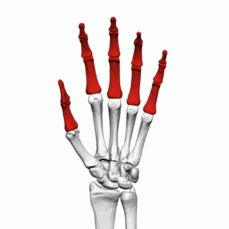
Phalangeal Fractures
Introduction[edit | edit source]
Fractures of the finger bones, known as phalanges, frequently occur and are often seen in emergency departments and clinics[1]. These injuries can affect the proximal, middle, or distal phalanx. In most cases of phalanx fractures, effective realignment can be achieved through non-surgical methods. Timely intervention is crucial to promote healing and restore functionality.
Clinical Anatomy[edit | edit source]
The hand's proximal and middle phalanges share a common anatomical structure comprising a head, neck, shaft, and base. Meanwhile, the distal phalanx is characterized by its tuft, shaft, and base divisions. The proximal phalanx is stabilized by surrounding structures, including proper and accessory collateral ligaments, the volar plate, and extensor/flexor tendons. The middle phalanx has two primary insertions: the central slip (part of the extensor mechanism) and the flexor digitorum superficialis (FDS). In the anatomy of the distal phalanx, the distal interphalangeal joint (DIPJ) is surrounded by extensor and flexor tendons, the volar plate, and collateral ligaments. The flexor digitorum profundus (FDP) inserts at the volar metaphysis of the distal phalanx. At the proximal interphalangeal joint (PIPJ), the flexor digitorum profundus and the flexor digitorum superficialis share a sheath. The flexor digitorum superficialis lies on the volar side, while the flexor digitorum profundus is on the dorsal side. As these tendons traverse the PIPJ, the flexor digitorum superficialis bifurcates into two slips, forming the Camper's chiasm, which inserts on the volar aspect of the middle phalanx. This significant anatomical relationship can result in a swan neck deformity, characterized by a hyperextended PIPJ and a flexed DIPJ.
Etiology[edit | edit source]
Phalangeal fractures of the hand are usually the result of a direct trauma, crush or twisting injury[2]
Epidemiology[edit | edit source]
Fractures involving the phalanges are prevalent and represent the most common injuries in the body. They are seen in athletic and work-related injuries[3]. Phalangeal fractures constitute around 18% of all upper-extremity fractures, making them the most prevalent fractures affecting the hand[4][5]. The majority of hand traumas involve the phalanges (46% phalangeal and 36% metacarpal). Among these, the distal phalanx and digits at the border are frequently affected. Males experience these injuries more frequently than females, and notably, the small finger is the most commonly injured
Classification of Phalangeal fractures[edit | edit source]
Fractures of the phalanx exhibit displacement patterns based on the level at which the fracture occurs, influenced by the intricate involvement of soft tissues and tendons.
Distal Phalanx[edit | edit source]
Fractures in the distal phalanx are typically nondisplaced or comminuted, falling into categories of tuft (tip), shaft, or articular injuries[6].
- Tuft Fractures: These commonly result from a crushing mechanism, such as striking the fingertip with a hammer. Tuft fractures often lead to open fractures, either due to associated soft tissue injury or nail bed involvement. Even without soft tissue damage, the presence of a nail bed injury classifies the fracture as open.
- Shaft Fractures: Intra-articular fractures are linked to extensor tendon avulsion (Mallet finger) or flexor digitorum profundus tendon avulsion.
- Mallet Finger: This involves the traumatic loss of terminal extension at the distal interphalangeal joint (DIPJ).
- Jersey Finger: Resulting from hyperextension, this injury involves avulsion of the flexor digitorum profundus.
- Seymour Fractures: Representing a displaced epiphyseal injury of the distal phalanx, often associated with nail bed injury, Seymour fractures typically result from hyperflexion and present as a mallet deformity with apex dorsal.
Middle Phalanx[edit | edit source]
Fractures in the middle phalanx lead to apex dorsal or volar angulation based on their location. Apex dorsal angulation occurs when the fracture is proximal to the flexor digitorum superficialis (FDS) insertion, causing displacement by the pull of the central slip. Apex volar angulation results from fractures distal to the FDS insertion. Fractures in the middle third may angulate in various directions or remain undisplaced due to the inherent stability provided by an intact and prolonged flexor digitorum superficialis insertion.
Proximal Phalanx[edit | edit source]
Fractures in the proximal phalanx exhibit apex volar angulation (dorsal angulation). The proximal fragment flexes due to interossei, while the distal phalanx extends as a result of the central slip.
Clinical Presentation[edit | edit source]
Patients with metacarpal fractures generally present with[7]
- Tenderness
- Swelling
- Bruising
- crepitus
- Deformity
- Restricted motion and instability
Diagnosis of Phalangeal fractures[edit | edit source]
Patients presentation : Individuals with phalangeal fractures typically seek attention at the Accident & Emergency (A&E) department following incidents like falls, sports-related injuries, or crush injuries to the hand[8]. During the initial presentation, it is essential to gather a comprehensive history, including details about the mechanism and timing of the injury, the extent and duration of any crushing force if applicable, and whether there is a disruption in the skin or a penetrating wound. A thorough understanding of the patient's medical and medication history is crucial, considering the potential impact of comorbidities (e.g., diabetes) or tobacco use on outcomes. Other significant aspects of the patient's history include hand dominance, occupation, and any hobbies involving manual dexterity.
Clinical examination of the hand is conducted by employing Apley's principles of 'look, feel, move,' with a comparison between the injured and uninjured hands[9]. Skin and soft-tissue changes, such as bruising, redness, wounds, asymmetry, or deformity, should be carefully observed. Any open wounds are examined to evaluate contamination levels and potential injuries to tendons, ligaments, or neurovascular structures. It is crucial to document examination findings, and clinical images can assist the Multidisciplinary Team (MDT) in their decision-making process. In conjunction with the fracture, there might be instances of shortening, angular deviations, or rotational deformities. Assessment of rotational malalignment involves observing the finger flexion cascade, illustrated in Figure 1, while the patient makes a fist. The alignment of the fingernail in relation to the palm and the nails of other digits should be parallel. The exclusion of malrotation is essential as it can impede hand function[10]. A comparative analysis with the uninjured hand is crucial, and any prior injuries to the same-side hand that could impact the flexion cascade should be considered.
A thorough neurovascular assessment is essential. Limbs should exhibit warmth and adequate perfusion, maintaining a capillary refill time within two seconds or less, and the skin temperature should be compared to the unaffected side[11]. Additionally, it is crucial to evaluate the intactness of motor and sensory functions in the radial, median, and ulnar nerves[12].
At the initial presentation, it is crucial to conduct a pain assessment. Various methods can be employed, including established tools like the visual analogue scale (VAS), which may be more suitable for children, or by simply asking the patient to rate their pain on a scale from 0 to 10, where 0 indicates no pain and 10 represents the most severe pain[13]. Regular assessments should be conducted based on pain evaluation
Potential diagnostic tests to consider include:
- Radiographs -Anteroposterior, lateral, and oblique images of each injured digit are needed. The lateral view should be taken so the other fingers do not obscure the injured digit. The oblique view can help diagnose fractures of the heads. Radiographs should be examined for angulation, rotational alignment, and shortening; the presence of intra-articular extension should also be noted. Rotational deformities are usually diagnosed via physical examination rather than radiographic finding.
- CT scan be useful if an intra-articular fracture is suspected but not seen on plain radiograp
Complications[edit | edit source]
Complications associated with phalangeal fractures can include[14][15][16]:
Delayed union
Nonunion: Usually of the atrophic type, however, it is an uncommon complication. There are various options of management including debridement, bone grafting, and plating, fusion, or ray amputation
Malunion:Either in the form of shortening, angulation, or malrotation. Where there is functional impairment as a result of malunion, surgical intervention would be indicated. This is either in the form of corrective osteotomy at the malunion site or metacarpal osteotomy that offers limited degrees of correction.
Soft tissue adhesions
Joint contractures:This is the most common complication. There is an increased risk of stiffness in phalanx fractures with prolonged immobilization, intra-articular extension, and where there is extensive dissection during operative management. Stiffness is managed with hand therapy rehabilitation and surgical release would be indicated only as a last resort of management.
Posttraumatic arthritis
Infection[17]
Management of Phalangeal fracture[edit | edit source]
The effective treatment of phalangeal fractures necessitates a comprehensive approach involving multiple disciplines(preoperative team, the intraoperative team and the postoperative team). The collaborative efforts of these various professionals are crucial for achieving the best possible restoration of hand function.
The initial evaluation should encompass a detailed medical history and clinical examination, supplemented by relevant radiological imaging, as these factors play a pivotal role in determining the suitable course of management. Once joint stabilization has been achieved to facilitate fracture union, prompt mobilization is essential to optimize the functional recovery of the hand. This review explores the principles of both operative and non-operative methods for managing such injuries.
Note: This article will concentrate on addressing the management of both closed and open fractures in patients.
Preoperative management of Phalangeals fractures[edit | edit source]
Initial management in accident and emergency[edit | edit source]
Preoperative pain management Initial pharmacological pain management involves prescribing non-opioid oral medications for milder pain and intravenous morphine for moderate-to-severe pain[18] . Careful medication management is necessary for patients with impaired breathing or cognition. Opiates should be avoided in individuals with concurrent head injuries[19] .
Conservative treatment[edit | edit source]
For stable fractures of phalangeal , nonsurgical management is the first-line therapy[20]
Two significant conservative treatment approaches are buddy taping and treatment using casts or splints.
Buddy Taping: Buddy taping is a straightforward, effective, and convenient method for treating certain phalangeal fractures. It is suitable for undisplaced fractures, impacted fractures, and distal phalanx tuft fractures. This technique involves securing the affected finger to an adjacent unaffected finger. Buddy taping allows for early range of motion exercises; however, there is a risk of stiffness developing in the unaffected finger.
Closed Reduction and Cast or Splint: This method is applied when fractures remain unstable even after closed reduction, such as in the case of unstable transverse fractures and dorsal fracture dislocation of the proximal interphalangeal (PIP) joint. It involves the use of a cast or splint for stabilization and support during the healing process.
Surgical treatment[edit | edit source]
In cases of unstable or complex phalangeal fractures, surgical stabilization offers advantages for long-term prognosis[21].Percutaneous pinning is recommended when fractures remain unstable following closed reduction. This method is suitable for various fracture types, including unstable transverse fractures, comminuted fractures, condylar fractures, and oblique/spiral fractures.
Open reduction and internal fixation are seldom necessary and are reserved for highly comminuted, oblique, or spiral fractures.
External fixators are utilized in cases of compound and highly comminuted fractures where open reduction and internal fixation are not feasible.
Physiotherapy measures in Phalangeal fractures[edit | edit source]
Early rehabilitation focuses on protecting the injuried phalanx. Intervention in this early period can include:
Patient education on edema control as it is an essential component of the initial therapy visit.Early mobilization to promote venous return viamuscle contraction is advocated in stable fractures.Having the patient adduct the fingers tightly and maintain this tension while flexing at the MP jointcan enhance both intrinsic muscle pumping and achieve the desired joint positions of full MP flexionand IP extension.Patients are also in-structed in shoulder and elbow ROM exercise in elevation to facilitate proximal muscle pumping.
Prescription or fabrication of a a protective orthosis(Splint) to provide support to a stable reduced fracture and properly provide safe resting position and to serially modify splints is one way to gradually allow a larger arc of motion over time.
Wound care (application of dressings or topical applications) are emphasized for edema control.
Swelling management (e.g. compression therapy, elevation or cryotherapy)
Scar management- Early controlled soft tissue and pain-modulated digital motion and tendon-gliding exercises are therefore given priority
Various workout routines are employed, frequently characterized by the specific exercises involved. The initiation of AROM (active range of motion) is prioritized at the earliest opportunity, determined by the fixation method, to hinder the formation of bony adhesions to tendons, ligaments, capsules, or skin. Among the crucial early tendon-gliding exercises are those targeting the flexor digitorum profundus (FDP), flexor digitorum superficialis (FDS), extensor digitorum communis, and the central slip, aiming to prevent tendon adherence to fracture callus
References[edit | edit source]
- ↑ Clarence Kee; Patrick Massey. Phalanx fracture. InStatPearls [Internet]2023 August 14. StatPearls Publishing.Available from:https://www.ncbi.nlm.nih.gov/books/NBK545182/
- ↑ Laura Kremer,Johannes Frank,Thomas Lustenberger,Ingo Marzi &Anna Lena Sander, Epidemiology and treatment of phalangeal fractures: conservative treatment is the predominant therapeutic concept, European Journal of Trauma and Emergency Surgery, Springer, Published online: 25 May 2020, volume 48, pages 567-571 (2022)
- ↑ J.J. de Jonge et al. Phalangeal fractures of the hand An analysis of gender and age-related incidence and aetiology J Hand Surg(1994)
- ↑ Karl JW, Olson PR, Rosenwasser MP (2015) The epidemiology of upper extremity fractures in the United States 2009. J OrthopTrauma 29(8):e242–e244.
- ↑ Onselen EBHV, Karim RB, Hage JJ (2003) Prevalence and dis- tribution of hand fractures. J Hand Surg Eur 28:491–495
- ↑ Schneider LH: Fractures of the distal phalanx. Hand Clin 1988;4:537-547.
- ↑ Benjamin J.F Dean, Christopher Little.Fractures of the metacarpals and phalanges.Orthopaedic and Trauma volume 25,Issue 1, February 2011,Pages 43-56
- ↑ BSSH 2022. General Information on Hand Fractures The British Society for Surgery of the Hand.
- ↑ Solomon L, Warwick D, Nayagam S, editors. Apley's system of orthopaedics and fractures. CRC press; 2010 Aug 27.
- ↑ Magee DJ, Zachazewski JE, Quillen WS, Manske RC. Pathology and intervention in musculoskeletal rehabilitation. Elsevier Health Sciences; 2015 Nov 20.
- ↑ Odak S, Bhalaik V. (i) Assessment of the acutely injured hand. Orthopaedics and Trauma. 2014 Aug 1;28(4):199-204.
- ↑ Thai JN, Pacheco JA, Margolis DS, Swartz T, Massey BZ, Guisto JA, Smith JL, Sheppard JE. Evidence-based comprehensive approach to forearm arterial laceration. Western Journal of Emergency Medicine. 2015 Dec;16(7):1127.
- ↑ Delgado DA, Lambert BS, Boutris N, McCulloch PC, Robbins AB, Moreno MR, Harris JD. Validation of Digital Visual Analog Scale Pain Scoring With a Traditional Paper-based Visual Analog Scale in Adults. J Am Acad Orthop Surg Glob Res Rev. 2018 Mar 23;2(3):e088
- ↑ Kurzen P, Fusetti C, Bonaccio M, et al. Complica- tions after plate fixation of phalangeal fractures. J Trauma 2006;60(4):841–3.
- ↑ Balaram AK, Bednar MS. Complications after the fractures of metacarpal and phalanges. Hand Clin 2010;26(2):169–77.
- ↑ Henry M. Hand fractures and dislocations. In: Rockwood CA, Green DP, Bucholz RW, editors. Rockwood and Green’s fractures in adults. 7th edi- tion. Philadelphia: Wolters Kluwer Health/Lippincott Williams & Wilkins; 2010. p. 710–829
- ↑ McLain, R. F., Steyers, C. & Stoddard, M. Infections in open fractures of the hand. The Journal of hand surgery 16, 108–112 (1991).
- ↑ National Institute for Health and Care Excellence (NICE) 2017. Fractures (complex): assessment and management (WWW document) Available at https://www.nice.org.uk/guidance/ng37 (Accessed 29 November 2023)
- ↑ National Institute for Health and Care Excellence (NICE) 2021. Co-codamol (WWW document) NICE Available at https://bnf.nice.org.uk/drug/co-codamol.html (Accessed 29 November 2021)
- ↑ Gaston, R. G. & Chadderdon, C. Phalangeal fractures: displaced/nondisplaced. Hand clinics 28, 395–401, x (2012).
- ↑ Fujioka, H., Takagi, Y., Tanaka, J. & Yoshiya, S. Corrective Step-cut Osteotomy at the Affected Bone for Correction of Rotational Deformity Due to Fracture of the Middle Phalanx. The journal of hand surgery Asian-Pacific volume 22, 240–243 (2017).


