
Overview of Neurodiagnostic Techniques
Original Editor - Stacy Schiurring
Top Contributors - Stacy Schiurring and Jess Bell
Introduction to Neurodiagnostics and Rehabilitation
Neurodiagnostics is a field of scientific study that involves studying and recording electrical activity in the central and peripheral nervous systems to help diagnose and treat neurological conditions.[1] Neuroimaging is the process of producing images of the structure or activity of the brain or other parts of the nervous system.[2]
While most rehabilitation professionals do not have the authority to directly request diagnostics or imaging, they frequently encounter imaging results through patient records or direct reports. Developing a strong understanding of neuroimaging and other neurodiagnostic techniques can significantly enhance a rehabilitation professional’s ability to evaluate and adapt treatment plans. It also fosters more effective collaboration with medical and nursing colleagues. Ultimately, this knowledge supports more informed clinical decisions, contributing to improved patient outcomes.
X-Ray Based Imaging
X-rays are a form of high-energy electromagnetic radiation used in medical imaging. They primarily visualise bones and joints, though they can also reveal some soft tissue structures. During an X-ray procedure, a controlled dose of radiation is directed through the body, and different tissues absorb the rays to varying degrees. Dense materials, like bones, absorb more X-rays and appear white on the resulting image, while less dense tissues, such as muscles and organs, absorb fewer rays and appear darker. The unabsorbed X-rays are captured on film or digital sensors, and the data is processed to produce a visual image of the body's internal structures.[3]

The nervous system does not show up clearly on standard X-rays, appearing in indistinct shades of grey due to its relatively low density.[3] To overcome this limitation, advanced imaging techniques, such as Computed Tomography and cerebral angiograms, have been developed to provide clearer and more detailed views of the nervous system.
Computed Tomography (CT)

Computed Tomography (CT) scanning is an advanced medical imaging technique that uses X-rays and computer processing to generate detailed cross-sectional images, or "slices," of the body. A CT scanner consists of a rotating X-ray tube and detectors that encircle the patient as they lie on a motorised table. As the scanner rotates, it sends X-rays through the body, and detectors measure how much radiation is absorbed by different tissues. Denser structures, like bones, absorb more X-rays and appear white, while softer tissues absorb less and appear darker. These measurements are compiled by a computer to create detailed internal images.
In neuroimaging, CT scans are essential for quickly evaluating brain injuries, strokes, tumours, and infections, especially in emergency settings. They are particularly effective in distinguishing between haemorrhagic and ischaemic strokes, which is a critical step in determining treatment. While CT scans are fast and widely available globally,[4] they offer less soft tissue contrast than magnetic resonance imaging (MRI, see below). This makes them less effective for analysing smaller brain structures or subtle abnormalities.[5]
CT scans can be tailored to specific body regions (e.g., a head or brain CT) and may be performed with or without contrast. A contrast agent enhances the visibility of blood vessels and certain tissues, making it easier to detect abnormalities like tumours, infections, and vascular conditions. These agents are typically injected intravenously, but can also be ingested or applied locally depending on the scan. While contrast agents improve diagnostic accuracy, they carry risks such as allergic reactions, kidney complications, and, rarely, contrast-induced nephropathy.[5] Therefore, a patient's medical history is reviewed before administration.

CT scanners are typically open-ended, allowing patients to see out both sides during the scan. This design helps reduce feelings of claustrophobia and anxiety, making the procedure more comfortable for many individuals.[5]
CT scans do expose patients to ionising radiation,[5] usually ranging from 1 to 10 millisieverts (mSv) per scan, depending on the body area. For context, the average person receives about 3 mSv of background radiation per year, and a chest X-ray delivers about 0.1 mSv. Though generally considered safe, this radiation exposure is a factor in deciding whether a CT scan is necessary, especially with repeated imaging.[6]
- Absolute contraindications to CT scanning: pregnancy (due to potential fetal radiation risks), severe allergic reactions to contrast materials, and pre- or mid-treatment for thyroid cancer with radioiodine, as contrast can interfere with therapy.[5][7]
- Relative contraindications: impaired kidney function, due to the risk of worsening renal problems; use of metformin, which raises the risk of lactic acidosis when combined with contrast; and conditions like hyperthyroidism, adrenal gland tumours, or myasthenia gravis.[7]
- Additionally, metal objects, such as implants, pacemakers, or surgical clips, may distort images and reduce their diagnostic quality.[7][8]
To learn more in-depth information about CT scans, please read this optional article.
Cerebral Angiography
Cerebral angiography is a specialised medical imaging procedure used to visualise the blood vessels in the brain and neck. It is commonly performed to diagnose vascular conditions such as aneurysms, arteriovenous (AV) malformations, stenosis, or blockages, and may also be used preoperatively to assess vascular health before brain or neck surgery. During the procedure, a catheter is inserted into a large blood vessel, typically in the groin or arm, and carefully guided through the vascular system to the arteries supplying the brain. A contrast agent is then injected through the catheter, allowing the blood vessels to be visualised on X-ray images, as the contrast absorbs X-rays differently from surrounding tissues. A series of real-time images is taken as the contrast flows through the cerebral vessels, producing highly detailed visualisations of the vascular structures.[9] [10]

Cerebral angiography is regarded as the gold standard for evaluating cerebral blood vessels due to its high resolution and ability to detect even small or subtle abnormalities. Although it is an invasive technique and carries some risks (e.g., bleeding, infection, and allergic reactions to the contrast agent), it remains a crucial diagnostic tool for serious neurological conditions.[9][10]
Positron Emission Tomography (PET)

Positron Emission Tomography (PET) is a minimally invasive imaging technique used to visualise metabolic and functional activity within the body. During a PET scan, a small amount of radioactive tracer is injected into the bloodstream, where it accumulates in tissues with high metabolic rates (e.g., tumours, areas of inflammation). As the tracer decays, it emits positrons that collide with nearby electrons, resulting in the emission of gamma rays. These gamma rays are detected by the PET scanner, where a computer processes the signals to produce images showing how the tracer is distributed throughout the body. PET scans are especially valuable for detecting cancer, monitoring brain activity, assessing cardiovascular health, and diagnosing neurological disorders, such as Alzheimer’s disease. Unlike imaging methods that only show anatomy, PET scans provide insight into the functional status of tissues and organs, making them a crucial tool for both diagnosis and treatment monitoring. While patients are exposed to a small amount of radiation from the tracer, the diagnostic benefits generally outweigh the risks.[11][12]
Often, PET scans are combined with CT scans in a procedure known as a PET/CT scan. This integrated approach merges functional imaging from PET scans with the detailed anatomical imaging of CT scans. While CT highlights the structure of tissues and organs, PET shows metabolic activity, allowing clinicians to see both where abnormalities are located and how actively they are functioning. For instance, in oncology, PET/CT can help identify malignant tumours based on elevated metabolic activity and determine their precise size and location. This combination enhances diagnostic accuracy and is particularly useful in cancer evaluation, neurological assessments, and cardiac imaging.[11][12]
- Contraindications to PET scanning: pregnancy, breastfeeding,[11] [12] immediately following or during chemotherapy or radiotherapy[13]
- Relative contraindications: patients with elevated blood glucose levels (especially above 200 mg/dL) may receive less reliable results,[12] and recent procedures like endoscopies, surgeries, or biopsies may impact the accuracy of PET imaging unless specifically cleared by the referring physician.[13]
Single-Photon Emission Computed Tomography (SPECT)

Single-Photon Emission Computed Tomography (SPECT) is a nuclear imaging technique used to produce detailed images of the body's internal physiological processes. Unlike CT scans, SPECT does not use X-rays. Instead, it involves the injection of a small amount of a radioactive tracer into the bloodstream. This tracer emits gamma rays, which are detected by a rotating gamma camera that moves around the patient. A computer then processes this data to create cross-sectional images showing how the tracer is distributed within the body. These images can highlight areas of abnormal function, such as reduced blood flow in the brain following a stroke or insufficient blood supply to parts of the heart in cases of coronary artery disease. SPECT is particularly valuable in the evaluation of heart conditions, neurological disorders, and some cancers. Because SPECT measures physiological function rather than anatomical structure, it is especially useful in diagnosing conditions, such as Parkinson’s disease, Alzheimer’s disease, seizure disorders, epilepsy, and dementia.[14]
SPECT is often combined with CT in a hybrid imaging procedure called SPECT/CT. This approach overlays functional data from the SPECT scan with the high-resolution anatomical images from the CT scan, improving diagnostic accuracy. The CT component helps to localise precisely where abnormal physiological activity is occurring, enabling clinicians to relate functional abnormalities to specific anatomical structures.[15]
- Contraindications for SPECT scanning: pregnancy, breastfeeding, recent myocardial infarction (within 2–4 days), acute myocarditis, acute pulmonary embolism, acute aortic dissection, severe symptomatic aortic stenosis, and severe pulmonary artery hypertension.[14][15]
- Relative contraindications: unstable angina, uncontrolled systemic hypertension, and untreated life-threatening arrhythmias.[14]
- These contraindications are related to the core function of SPECT in evaluating blood flow and cardiovascular function, where acute or unstable pathologies can complicate image accuracy or patient safety.
Magnetic Resonance Imaging (MRI)

Magnetic Resonance Imaging (MRI) is a non-invasive medical imaging technique that uses strong magnetic fields, radiofrequency pulses, and advanced computer processing to produce detailed images of the body’s internal structures. The process begins by aligning hydrogen atoms, which are abundant in water and fat, within the body using a powerful magnet. A radiofrequency pulse is then applied to temporarily disturb this alignment. As the atoms return to their original positions, they emit signals that vary depending on the type of tissue. These signals are detected by specialised coils and converted by a computer into highly detailed cross-sectional images, which can be viewed from multiple angles. Unlike CT scans, MRI does not involve ionising radiation. This makes MRI a safer option, particularly for visualising soft tissues such as the brain, spinal cord, muscles, and internal organs. MRI is particularly effective in detecting abnormalities like tumours, inflammation, and degenerative diseases.[16]
In some cases, contrast agents are administered intravenously to enhance image clarity. These agents alter the magnetic properties of certain tissues, making abnormal areas such as tumours or inflamed regions more visible on the scan. Contrast is used in roughly one-third of MRI procedures and is generally safe, though it should be avoided in patients with specific kidney conditions or known allergies to contrast ingredients.[16][17]
MRI technology plays a vital role in both clinical diagnostics and research. A single scanner can be used for various types of scans,[17] depending on the imaging protocol and the anatomical region being studied.
| Structures imaged and /or information provided | |
|---|---|
| General MRI | Creates detailed images of the entirety of a person's anatomy |
| Structural MRI | Focuses on creating images of the anatomy and structure of the central nervous system (CNS) |
| Functional MRI (fMRI)[18] | Measures and examines brain activity and function by detecting changes in blood flow related to neural activity |
| Diffusion Tensor Imaging (DTI)[19] | Provides information about the structural organisation and integrity of white matter tracts |
| Magnetic Resonance Angiography (MRA)[20] | Creates high-resolution images of blood vessels, allowing for the assessment of conditions like aneurysms, stenosis, or blockages |
-
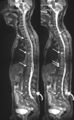 Example of MRI image
-
 Example of functional MRI image
-
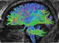 Example of Diffusion Tensor Imaging
-
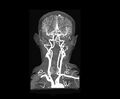 Example of Magnetic Resonance Angiography


Access to MRI is highly unequal across the globe. High-income countries and urban areas typically have more scanners, whereas low- and middle-income countries and rural regions often lack adequate access. It is estimated that approximately two-thirds of the global population do not have access to MRI scanning.[21]
While MRI is generally considered safe, certain risks and contraindications must be considered. Patients with metal implants (e.g., pacemakers, cochlear implants, insulin pumps, surgical clips, or embedded shrapnel) may be at risk due to the strong magnetic field. Even items like magnetic dental implants, some piercings, or older tattoos containing metal-based inks can pose safety concerns. Although many medical implants are now manufactured to be MRI-compatible, all devices must be carefully screened before scanning. MRI is also typically avoided during the first trimester of pregnancy unless medically necessary, as the long-term effects of magnetic fields on fetal development are not fully known. Additionally, patients with claustrophobia or anxiety may find traditional closed MRI machines uncomfortable.[17][22] In such cases, open MRI machines, which are less confining, are increasingly available and can provide a more tolerable alternative.
To learn more in-depth information about MRI scans, please read this optional article.
Electroencephalography (EEG)

Electroencephalography (EEG) is a diagnostic method used to measure and record the brain's electrical activity. The procedure involves placing small electrodes on the scalp, which detect the electrical impulses generated as neurons communicate with one another. These signals are then amplified and recorded by a machine, typically displayed as brainwave patterns on a computer screen or printout. The recorded brainwaves are categorised into frequency bands (e.g., alpha, beta, delta, and theta waves); each band is associated with different arousal and mental states.[23]
It is a non-invasive, safe, and painless procedure that provides real-time insight into brain function. EEG allows clinicians to directly observe the brain's electrical activity. This plays a crucial role in both clinical diagnosis and research into brain-related disorders. EEG is most commonly used to diagnose neurological conditions, such as epilepsy, sleep disorders, and brain injuries, as it can reveal abnormal electrical activity, like seizures or irregular brainwave patterns.[23]
Nerve Conduction Study

A nerve conduction study is a diagnostic test used to evaluate the function of the peripheral nerves. During the procedure, small electrodes are placed on the skin over the nerve being examined: one electrode delivers a mild electrical impulse to stimulate the nerve, while another measures the response as the signal travels along the nerve pathway. This test assesses the speed (conduction velocity) and strength (amplitude) of the nerve's electrical signal, providing critical information about the health of the nerve fibres. It is particularly useful in diagnosing conditions such as carpal tunnel syndrome, peripheral neuropathy, and other nerve-related disorders. The presence of delays or blockages in signal transmission can help pinpoint the type and location of nerve damage. While the procedure is generally safe and well tolerated, some individuals may experience mild discomfort or tingling during stimulation.[24]
Nerve conduction studies are especially valuable in physical rehabilitation settings, where they support the assessment of nerve and muscle function. By identifying nerve damage or dysfunction, these studies inform clinicians about the severity and nature of the condition, helping to guide treatment planning. They also provide a means of tracking progress, as improvements in nerve conduction can indicate recovery over time. Additionally, the objective data gained from these tests helps therapists develop targeted rehabilitation programmes that address deficits in strength, coordination, or sensation.[24]
To learn more in-depth information about nerve conduction studies, please read this optional article.
Resources
Optional Additional Reading
- Boyles RE, Gorman I, Pinto D, Ross MD. Physical therapist practice and the role of diagnostic imaging. journal of orthopaedic & sports physical therapy. 2011 Nov;41(11):829-37.
- Keil AP, Hazle C, Maurer A, Kittleson C, Watson D, Young B, et al. Referral for imaging in physical therapist practice: key recommendations for successful implementation. Physical Therapy. 2021 Mar 1;101(3):pzab013.
Clinical Resource
- Radiology Masterclass: MRI interpretation (for educational purposes only, always refer to a Radiologist for imaging interpretation)
References
- ↑ Neurodiagnostic Society. Neurodiagnostics. Available from: https://www.aset.org/neurodiagnostics/ (accessed 19 May 2025).
- ↑ Oxford Languages. Neuroimaging. Available from: https://languages.oup.com/google-dictionary-en/(accessed 19 May 2025).
- ↑ 3.0 3.1 NIH National Institute of Biomedical Imaging and Bioengineering. X-rays. Available from: https://www.nibib.nih.gov/science-education/science-topics/x-rays (accessed 18 May 2025).
- ↑ Collective Minds. Number of CT Scans Per Year Worldwide: Overview of global Computer Tomography utilization. Available from: https://about.cmrad.com/articles/number-of-ct-scans-per-year-worldwide-overview-of-global-computer-tomography-utilization (accessed 18 May 2025).
- ↑ 5.0 5.1 5.2 5.3 5.4 NIH National Institute of Biomedical Imaging and Bioengineering. Computed Tomography (CT). Available from: https://www.nibib.nih.gov/science-education/science-topics/computed-tomography-ct (accessed 18 May 2025).
- ↑ National Cancer Institute. Computed Tomography (CT) Scans and Cancer. Available from: https://www.cancer.gov/about-cancer/diagnosis-staging/ct-scans-fact-sheet (accessed 18 May 2025).
- ↑ 7.0 7.1 7.2 Rawson JV, Pelletier AL. When to order contrast-enhanced CT. American family physician. 2013 Sep 1;88(5):312-6.
- ↑ Patel P, De Jesus O. CT Scan [Internet]. 2023 [cited 18 May 2025]. Available from: https://www.ncbi.nlm.nih.gov/books/NBK567796/
- ↑ 9.0 9.1 RadiologyInfo. Cerebral Angiography. Available from: https://www.radiologyinfo.org/en/info/angiocerebral (accessed 19 May 2025).
- ↑ 10.0 10.1 Cleveland Clinic. Cerebral Angiogram. Available from: https://my.clevelandclinic.org/health/diagnostics/13476-cerebral-angiogram (accessed 19 May 2025).
- ↑ 11.0 11.1 11.2 Mayo Clinic. Positron Emission Tomography Scan. Available from: https://www.mayoclinic.org/tests-procedures/pet-scan/about/pac-20385078 (accessed 18 May 2025).
- ↑ 12.0 12.1 12.2 12.3 Cleveland Clinic. PET Scan. Available from: https://my.clevelandclinic.org/health/diagnostics/10123-pet-scan (accessed 18 May 2025).
- ↑ 13.0 13.1 United Imaging. PET/CT scan. Available from: https://eu.united-imaging.com/en/news-center/knowledge-base/category-ct/article-7 (accessed 18 May 2025).
- ↑ 14.0 14.1 14.2 Yandrapalli S, Puckett S. SPECT Imaging [Internet]. 2022 [cited 19 May 2025]. Available from: https://www.ncbi.nlm.nih.gov/books/NBK564426/
- ↑ 15.0 15.1 Cleveland Clinic. SPECT Scan. Available from: https://my.clevelandclinic.org/health/diagnostics/spect-scan (accessed 19 May 2025).
- ↑ 16.0 16.1 NIH National Institute of Biomedical Imaging and Bioengineering. Magnetic Resonance Imaging (MRI). Available from:https://www.nibib.nih.gov/science-education/science-topics/magnetic-resonance-imaging-mri (accessed 19 May 2025).
- ↑ 17.0 17.1 17.2 Ashby K, Adams B, Shetty M. Appropriate Magnetic Resonance Imaging Ordering [Internet]. 2022 [cited 19 May 2025]. Available from: https://www.ncbi.nlm.nih.gov/books/NBK565857/
- ↑ Glover GH. Overview of functional magnetic resonance imaging. Neurosurgery Clinics of North America. 2011 Apr;22(2):133.
- ↑ Ranzenberger L, Das J, Snyder T. Diffusion Tensor Imaging [Internet]. 2023 [cited 19 May 2025]. Available from: https://www.ncbi.nlm.nih.gov/books/NBK537361/
- ↑ RadiologyInfo. MR Angiography (MRA). Available from: https://www.radiologyinfo.org/en/info/angiomr(accessed 19 May 2025).
- ↑ Jalloul M, Miranda‐Schaeubinger M, Noor AM, Stein JM, Amiruddin R, Derbew HM, Mango VL, Akinola A, Hart K, Weygand J, Pollack E. MRI scarcity in low‐and middle‐income countries. NMR in Biomedicine. 2023 Dec;36(12):e5022.
- ↑ Mayo Clinic. MRI. Available from: https://www.mayoclinic.org/tests-procedures/mri/about/pac-20384768 (accessed 19 May 2025).
- ↑ 23.0 23.1 NHS. Electroencephalogram (EEG). Available from: https://www.nhs.uk/conditions/electroencephalogram/ (accessed 19 May 2025).
- ↑ 24.0 24.1 Tavee J. Nerve conduction studies: Basic concepts. Handbook of clinical neurology. 2019 Jan 1;160:217-24.