File:TDCS.png
TDCS.png (264 × 237 pixels, file size: 140 KB, MIME type: image/png)
Summary[edit | edit source]
https://commons.wikimedia.org/wiki/File:TDCS_administration.gif
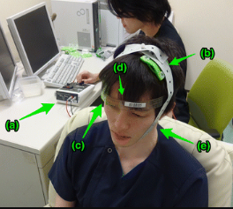
tDCS administration at National Center of Neurology and Psychiatry Hospital. A subject (front) sits on a sofa relaxed, and a researcher (behind) controls the tDCS device (a). In this picture, anodal (b) and cathodal (c) electrodes with 35-cm2 size are put on F3 and right supraorbital region, respectively. We use a head strap (d) for convenience and reproducibility, and also use a rubber band (e) for reducing resistance.
Licensing[edit | edit source]
This file is licensed under the Creative Commons Attribution-ShareAlike 4.0 International.
You are free to:
- Share - copy and redistribute the material in any medium or format
- Adapt - remix, transform, and build upon the material for any purpose, even commercially.
The licensor cannot revoke these freedoms as long as you follow the license terms.
Under the following terms:
- Attribution – You must give appropriate credit, provide a link to the license, and indicate if changes were made. You may do so in any reasonable manner, but not in any way that suggests the licensor endorses you or your use.
- ShareAlike - If you remix, transform or build upon the material, you must distribute your contributions under the same licence as the original.
No additional restrictions — You may not apply legal terms or technological measures that legally restrict others from doing anything the license permits.
File history
Click on a date/time to view the file as it appeared at that time.
| Date/Time | Thumbnail | Dimensions | User | Comment | |
|---|---|---|---|---|---|
| current | 16:08, 9 December 2023 | | 264 × 237 (140 KB) | Angeliki Chorti (talk | contribs) | https://commons.wikimedia.org/wiki/File:TDCS_administration.gif tDCS administration at National Center of Neurology and Psychiatry Hospital. A subject (front) sits on a sofa relaxed, and a researcher (behind) controls the tDCS device (a). In this picture, anodal (b) and cathodal (c) electrodes with 35-cm2 size are put on F3 and right supraorbital region, respectively. We use a head strap (d) for convenience and reproducibility, and also use a rubber band (e) for reducing resistance. |
You cannot overwrite this file.
File usage
The following page uses this file:

