File:Insertion site of ACL on Femur.jpg
Original file (2,007 × 1,134 pixels, file size: 209 KB, MIME type: image/jpeg)
Summary[edit | edit source]
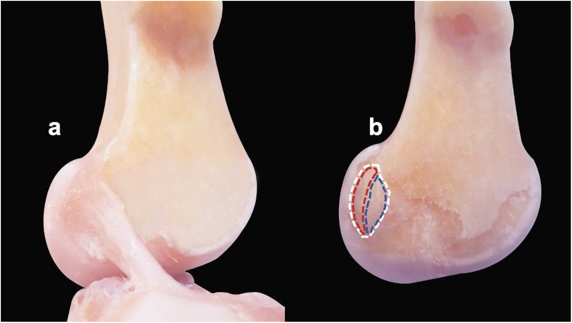
Femoral insertion sites of the ACL in the specimens before and after the ligament is cut. (a) ACL with an oval/elliptical morphology with its intact tibial and femoral attachments. (b) ACL removed showing the femoral footprint (white dashed line) with its anterior border formed by the lateral intercondylar ridge (resident's ridge) and posterior articular margin of the lateral femoral condyle forming its posterior border in continuity with the posterior femoral cortex. Dashed blue line marking direct insertion and red dashed line representing indirect fan-like insertion.
Avalos RM, Torres-González EM, Padilla-Medina JR, Monllau JC. ACL ANATOMY: IS THERE STILL SOMETHING TO LEARN?. Revista Española de Cirugía Ortopédica y Traumatología. 2023 Feb 12.
Licensing[edit | edit source]
File history
Click on a date/time to view the file as it appeared at that time.
| Date/Time | Thumbnail | Dimensions | User | Comment | |
|---|---|---|---|---|---|
| current | 14:56, 20 July 2023 | | 2,007 × 1,134 (209 KB) | Rujuta Naik (talk | contribs) | Femoral insertion sites of the ACL in the specimens before and after the ligament is cut. (a) ACL with an oval/elliptical morphology with its intact tibial and femoral attachments. (b) ACL removed showing the femoral footprint (white dashed line) with its anterior border formed by the lateral intercondylar ridge (resident's ridge) and posterior articular margin of the lateral femoral condyle forming its posterior border in continuity with the posterior femoral cortex. Dashed blue line marking... |
You cannot overwrite this file.
File usage
The following page uses this file:

