File:Anterior view of posterolateral attachments of lateral menicus.jpeg

Size of this preview: 673 × 599 pixels. Other resolution: 1,890 × 1,683 pixels.
Original file (1,890 × 1,683 pixels, file size: 608 KB, MIME type: image/jpeg)
Summary[edit | edit source]
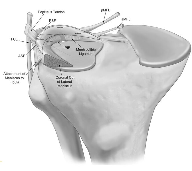
Anterior view illustration of the anatomic attachments to the posterolateral meniscus. The meniscotibial ligament and the posteroinferior popliteomeniscal fascicle (PIF) are depicted emerging from posterior to the lateral tibial plateau and attaching to the inferior margin of the posterolateral meniscus. aMFL, anterior meniscofemoral ligament; pMFL, posterior meniscofemoral ligament; ASF, anterosuperior popliteomeniscal fascicle; PSF, posterosuperior popliteomeniscal fascicle; FCL, fibular collateral ligament.[1]
Licensing[edit | edit source]
The copyright holder has given special permission to use this work in Physiopedia.
- ↑ Aman ZS, DePhillipo NN, Storaci HW, Moatshe G, Chahla J, Engebretsen L, et al. Quantitative and qualitative assessment of posterolateral meniscal anatomy: Defining the popliteal hiatus, popliteomeniscal fascicles, and the lateral meniscotibial ligament. Am J Sports Med [Internet]. 2019;47(8):1797–803. Available from: http://dx.doi.org/10.1177/0363546519849933
File history
Click on a date/time to view the file as it appeared at that time.
| Date/Time | Thumbnail | Dimensions | User | Comment | |
|---|---|---|---|---|---|
| current | 17:03, 31 March 2023 | | 1,890 × 1,683 (608 KB) | Robert Pierce (talk | contribs) | Anterior view illustration of the anatomic attach- ments to the posterolateral meniscus. The meniscotibial liga- ment and the posteroinferior popliteomeniscal fascicle (PIF) are depicted emerging from posterior to the lateral tibial pla- teau and attaching to the inferior margin of the posterolateral meniscus. aMFL, anterior meniscofemoral ligament; pMFL, posterior meniscofemoral ligament; ASF, anterosuperior popliteomeniscal fascicle; PSF, posterosuperior popliteome- niscal fascicle; FCL, fi... |
You cannot overwrite this file.
File usage
The following page uses this file:

