Cleidocranial Dysplasia (CCD)
Original Editor - Ravi Kumar
Top Contributors - Ravi Kumar
Introduction[edit | edit source]
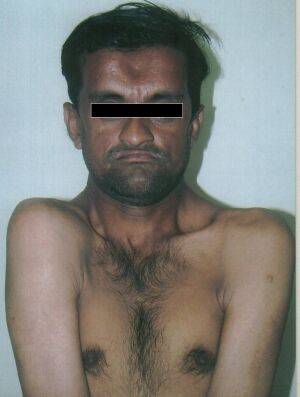
Cleidocranial Dysplasia (CCD) is a rare genetic disorder that affects the development and growth of teeth and bones such as the skull, face, spine, collarbones, and legs[1]. The name “cleidocranial dysplasia” comes from “cleido,” which refers to the collarbones, and “cranial,” which refers to the skull[2]. It is also known as Scheuthauer- Marie-Sainton syndrome[3], People with CCD have abnormal or missing collarbones, which allow them to bring their shoulders close together in front of their chest[4]. They also have delayed closure of the gaps between the bones of the skull, which results in a large and prominent forehead[4]. CCD can cause various dental problems, such as delayed eruption of permanent teeth, extra teeth, or poorly aligned teeth. Some people with CCD may also have other skeletal abnormalities, such as short stature, curved spine, or malformed pelvis.
CCD is either inherited or caused by mutations in the RUNX2 gene,[5] a gene required for osteoblastic differentiation, which is involved in the formation of bone and cartilage cells. The condition is inherited in an autosomal dominant manner, which means that one copy of the mutated gene is enough to cause the disorder. However, some cases of CCD occur randomly due to new mutations in the gene. The severity and features of CCD can vary widely among affected individuals, even within the same family.
Epidemiology[edit | edit source]
Cleidocranial dysplasia (CCD) is a rare genetic disorder that affects the development of bones and teeth. It is estimated to have a prevalence of approximately 1 in 1,000,000 individuals worldwide[7].
It is considered a rare or orphan disease within the group of primary bone dysplasias.[8]
The disorder is found in many ethnic groups, and no sex predilection has been reported. It may be underdiagnosed because of the number of relatively mild cases[9].
Signs and symptoms[edit | edit source]
Symptoms of CCD can vary widely in severity, even within the same family. The most common symptoms of CCD include[10]:
- Abnormal dental enamel morphology
- Abnormality of the dentition
- Carious teeth
- Down-sloping shoulders
- Frontal bossing
- High, narrow palate
- Hypertelorism
- Hypoplasia of the zygomatic bone
- Hypoplastic inferior ilia
- Large fontanelles
- Micrognathia
- Narrow chest
- Recurrent respiratory infections
- Short clavicles
- Short stature
- Skeletal dysplasia
- Sloping forehead
- Hearing impairment
- Osteoporosis
Diagnosis[edit | edit source]
Diagnosis of CCD is based on clinical signs and characteristic radiographic findings. These findings encompass the presence of wide-open sutures, patent fontanels, cone-shaped thorax with narrow upper thoracic diameter, hand deformities, abnormal dentition. Molecular genetic testing of RUNX2 gene can be used to confirm the diagnosis in patients with atypical clinical and radiological diagnostic features.
Treatment[edit | edit source]
There is no cure for CCD, but the symptoms can be managed with various treatments depending on the individual's needs. Some of the possible treatments are:-
Craniofacial surgery[11]
Spinal fusion procedures to stabilize the spine and prevent scoliosis[12]
Removal of collarbone fragments to improve shoulder mobility
Helmets to protect children against brain injury until their fontanels close
Protective equipment to prevent injuries, particularly while playing sports
Calcium and vitamin D supplements to strengthen bones
Dental procedures to remove extra teeth, align crowded teeth and replace missing teeth
Treatment of sinus and ear infections to prevent hearing loss
References[edit | edit source]
- ↑ Kolokitha OE, Ioannidou I. A 13-year-old Caucasian boy with cleidocranial dysplasia: a case report. BMC research notes. 2013 Dec;6:1-6.
- ↑ Ickow IM. Cleidocranial dysplasia (CCD) [Internet]. 2021. Available from: https://www.hopkinsmedicine.org/health/conditions-and-diseases/cleidocranial-dysplasia-ccd
- ↑ Kuruvila VE, Bilahari N, Attokkaran G, Kumari B. Scheuthauer-Marie-Sainton syndrome. Contemporary Clinical Dentistry. 2012 Jul;3(3):338.
- ↑ 4.0 4.1 Cleidocranial dysplasia: Medlineplus Genetics [Internet]. U.S. National Library of Medicine;
- ↑ Dalle Carbonare L, Antoniazzi F, Gandini A, Orsi S, Bertacco J, Li Vigni V, Minoia A, Griggio F, Perduca M, Mottes M, Valenti MT. Two Novel C-Terminus RUNX2 Mutations in Two Cleidocranial Dysplasia (CCD) Patients Impairing p53 Expression. International Journal of Molecular Sciences. 2021 Sep 25;22(19):10336.
- ↑ Air to air. Cleidocranial dysplasia Available from: https://www.youtube.com/watch?v=6L2qRi-cyCE[last accessed 16/04/2023]
- ↑ Cano-Pérez E, Gómez-Alegría C, Herrera FP, Gómez-Camargo D, Malambo-García D. Demographic, clinical, and radiological characteristics of cleidocranial dysplasia: A systematic review of cases reported in south America. Annals of Medicine and Surgery. 2022 Apr 10:103611.
- ↑ Segovia‐Fuentes JI, Egurrola‐Pedraza JA, Castro‐Mendoza EJ, Cano‐Pérez E, Gómez‐Camargo DE, Malambo‐García DI. Clinical‐radiological approach for the diagnosis of cleidocranial dysplasia in adults: A familial cases series. Clinical Case Reports. 2021 Dec;9(12):e05235.
- ↑ INSERM US14 -- ALL RIGHTS RESERVED [Internet]. . Available from: https://www.orpha.net/consor/cgi-bin/OC_Exp.php?Expert=1452&Lng=GB
- ↑ Cleidocranial dysplasia - about the disease [Internet]. U.S. Department of Health and Human Services; [cited 2023 Jun 10]. Available from: https://rarediseases.info.nih.gov/diseases/6118/cleidocranial-dysplasia
- ↑ Jirapinyo C, Deraje V, Huang G, Gue S, Anderson PJ, Moore MH. Cleidocranial dysplasia: management of the multiple craniofacial and skeletal anomalies. Journal of Craniofacial Surgery. 2020 Jun 1;31(4):908-11.
- ↑ Balioğlu MB, Kargın D, Albayrak A, Atıcı Y. The treatment of cleidocranial dysostosis (Scheuthauer-Marie-Sainton Syndrome), a rare form of skeletal dysplasia, accompanied by spinal deformities: a review of the literature and two case reports. Case Reports in Orthopedics. 2018 Jul 9;2018.


