When refering to evidence in academic writing, you should always try to reference the primary (original) source. That is usually the journal article where the information was first stated. In most cases Physiopedia articles are a secondary source and so should not be used as references. Physiopedia articles are best used to find the original sources of information (see the references list at the bottom of the article).
If you believe that this Physiopedia article is the primary source for the information you are refering to, you can use the button below to access a related citation statement.
Boerhaave syndrome typically occurs after forceful emesis and retching. Boerhaave syndrome is a transmural perforation of the esophagus and should be distinguished from Mallory-Weiss syndrome, a nontransmural esophageal tear also associated with vomiting. Since the perforation occurs with emesis, Boerhaave syndrome is usually not truly spontaneous, but this term helps distinguish it from iatrogenic perforation. Vomiting is the most common cause, but any activity that increases intraesophageal pressure can result in this syndrome. This condition can manifest in patients with a typically functioning esophagus, yet there is a subgroup where specific esophageal abnormalities or pathology are identified. Boerhaave syndrome accounts for 10% to 15% of all esophageal perforations.
Diagnosis of this condition can be challenging because the presentation can vary significantly, thus requiring an index of suspicion. Boerhaave syndrome is classically associated with the Mackler triad of vomiting, chest pain, and subcutaneous emphysema. However, patients rarely present with all of these symptoms and often have vague, nonspecific complaints. This can contribute to a delay in diagnosis and poor outcomes. Boerhaave syndrome is one of the most lethal gastrointestinal tract disorders, with a mortality rate of up to 60% with intervention, increasing to nearly 100% without intervention.[2] Treatment is varied and depends on the time of diagnosis and the patient’s clinical condition at presentation. Management can range from conservative management to major surgical resection.[1]
Etiology
Boerhaave syndrome is a barogenic injury resulting from a sudden increase in intraluminal pressure against a closed cricopharyngeus. Neuromuscular dysfunction results in a non-relaxed cricopharyngeus with a resultant rise in pressure. This pressure overwhelms the wall of the esophagus at its weakest point.[4] In adults, the perforation commonly occurs in the left posterolateral aspect of the distal esophagus below the diaphragm; however, in very young patients, the esophagus usually perforates into the right pleural cavity.
Common risk factors include alcoholism and overindulgence in food. Both can lead to sudden forceful emesis, usually the most common cause. Other causes that raise pressure within the esophagus include weightlifting, defecation, epileptic seizures, abdominal trauma, compressed air injury, and childbirth.[5] Most cases occur in patients with a normal underlying esophagus, although esophagitis and ulcers have also been found in a subset of individuals. No apparent genetic predisposition exists for Boerhaave syndrome.[1]
Evaluation
The evaluation consists mainly of physical examination and radiographic assessment. A focused physical examination is essential to ascertain the degree of distress or physiologic derangement.
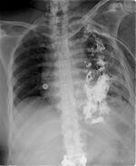
xrayImaging is critical in diagnosing Boerhaave syndrome. Plain films of the chest and abdomen may show subcutaneous or mediastinal emphysema, mediastinal widening, and pleural effusion. In up to 20% of cases, the Nacleario V-sign may be seen as radiolucent streaks that dissect the retrocardiac fascia to form the letter V. This is a specific but insensitive radiographic sign of esophageal perforation.
Laboratory tests are often nonspecific in the diagnosis of Boerhaave syndrome. Patients may have a leukocytosis with a left shift, and many have a hematocrit value of around 50% due to hemoconcentration from fluid loss. Sampling from a present pleural effusion may show undigested food particles and gastric fluid. Lab results usually reveal a pH of less than 6 and an elevated salivary amylase level.
A contrast esophagogram may aid in diagnosis. A water-soluble contrast agent such as Gastrografin should be used since extravasation of barium can lead to mediastinitis and subsequent fibrosis. The sensitivity of this study is dependent upon the size and location of the perforation and technique. False-negative results occur in 10% to 38% of cases. The study may be repeated with barium if the initial test with water-soluble contrast is negative.
CT imaging aids in a more definitive diagnosis and may be better tolerated in severely ill patients. CT imaging provides more detail regarding the location of drainable collections and aids in localizing the rupture site. A contrast medium may further define the injury extent and assist in a timely diagnosis. CT findings may include periesophageal and mediastinal gas, mediastinal fluid collections, esophageal wall thickening, pleural effusion, pneumothorax, and hydrothorax.
Endoscopy is considered controversial as it increases the risk of further esophageal perforation. Endoscopy should be reserved for patients whose perforation location is unclear and who are appropriate candidates for endoscopic treatment.[1]
Differential diagnosis
Symptoms of Boerhaave syndrome are often nonspecific and may be seen with many other conditions such as aortic dissection, pancreatitis, myocardial infarction, pulmonary embolus, perforated peptic ulcer, spontaneous pneumothorax, pneumonia, pericarditis, or Mallory-Weiss tear. These disorders can be distinguished from Boerhaave syndrome by history, physical examination, laboratory evaluation, electrocardiogram, and other imaging.[1]
Management
The 3 common treatment modalities for Boerhaave syndrome include nonsurgical, endoscopic, and surgical (open vs minimally invasive). The management decision is best made by an interprofessional team with experience in all treatment options. Treatment often consists of a combination of medical and surgical interventions. The mainstay of treatment includes avoidance of all oral intake, volume replacement, broad-spectrum antibiotic coverage, nutritional support (usually parenteral), source control of any leaks (eg, via IR or VATS), and prompt surgical or endoscopic intervention as indicated. Treatment approaches are typically customized based on the perforation's location and size, the duration between injury and diagnosis, and the patient's overall condition. Early diagnosis (within 12 to 24 hours) yields the best outcome.
Criteria for patients who are candidates for medical management include the following:
The leak is contained within the neck or mediastinum or between the mediastinum and visceral lung pleura.
Contrast can flow back into the esophagus from the cavity surrounding the perforation.
The injury is not in neoplastic tissue, in the abdomen, or proximal to an obstruction.
The patient has minimal symptoms with no signs of sepsis.
Contrast studies can be obtained at any time of day.
Surgical specialists are readily available if the patient deteriorates.
Medical management includes avoidance of all oral intake for at least 7 days, parenteral nutrition, broad-spectrum IV antibiotics for 7 to 14 days, and drainage of any fluid collections. Patients who show signs of clinical deterioration during conservative treatment require surgical intervention. Surgery is indicated if any of the following develops: contained perforation develops into a free perforation, the injury extends, the patient exhibits persistent fevers, clinical deterioration including the development of sepsis, progression of pneumothorax/pneumomediastinum, or development of empyema. The most successful surgical method often entails primary esophageal repair, particularly within the first 4 hours of perforation. This can be achieved through open thoracotomy vs VATS with fundic reinforcement, which is now considered the gold standard of treatment.
In a diseased, nonviable segment of the esophagus, resection may be the best treatment. In late presentations diagnosed after 24 hours, the wound edges are typically edematous, stiff, or friable, rendering primary repair risky. Considering this, many manage late perforations with debridement of the pleural cavity and mediastinum, esophagostomy, and feeding gastrostomy. Definitive reconstruction may be performed after 6 weeks. Although surgery is the most common treatment of Boerhaave syndrome, stenting across the leak has been used with promising results in some instances. This option might be considered in patients with significant underlying comorbidities. Advanced endoscopic techniques can provide patients with a minimally invasive therapeutic intervention. Endoscopic approaches for managing esophageal perforation include placement of fully covered esophageal stents, through-the-scope clips, over-the-scope clips, and endoscopic suturing, esophageal resection and diversion.[1]