Wound Healing
Original Editor Esraa Mohamed Abdullzaher
Top Contributors - Esraa Mohamed Abdullzaher, Naomi O'Reilly, Lucinda hampton, Kim Jackson, Olajumoke Ogunleye, Joao Costa, Claire Knott, Aya Alhindi, Admin and Lauren Lopez
Anatomy and function of the skin[edit | edit source]
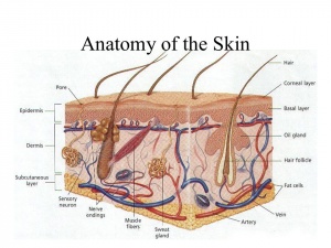
Skin is the largest organ in the body and performs a wide variety of different functions. It is composed of a predominantly cellular epidermis and an underlying dermis, which is composed of fibers of connective tissue relatively sparsely populated with cells . They play an important role in the injury of the skin.
The epidermis contains mostly keratinocytes, among which are interspersed melanocytes, Merkel cells, Langerhans cells, and other resident immune cells. Keratinocytes are responsible for the production of keratin, a fibrous structural protein that contributes to the strength and waterproofing of skin. Melanocytes protect against ultraviolet (UV) light, producing melanin, the dark pigment that gives the skin its color. Langerhans cells are professional antigen-presenting cells and play critical roles in both protective immune responses in the skin and maintenance of immune homeostasis. Merkel cells have been associated with discrimination of light touch.The epidermis also contains dermal appendages, such as hair follicles, sebaceous glands, eccrine sweat glands, and apocrine glands.
The dermis is the layer between the epidermis and the subcutaneous tissue. It is made up of collagen, elastic fibers, and an extrafibrillar matrix.The dermis is a highly vascularized structure . the deepest of which is the fascial plexus, located at the level of the deep muscle fascia. Overlying this, at the level of the superficial fascia, is the subcutaneous plexus. The next level is the subdermal plexus, an extensive vascular network situated at the junction between the reticular dermis and the subcutaneous tissue directly below, which has an important role in the distribution of blood to other regions of the cutaneous system. Superficial to the subdermal plexus is the subpapillary plexus, situated between the papillary and reticular dermis with capillary loops extending into the papillae. Blood flow between the subdermal and subpapillary plexus is achieved through a series of arterioles and venules and has an important role in regulation of body temperature and the metabolic supply of the skin.
Types of wound[edit | edit source]
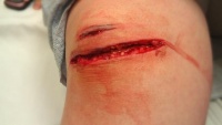
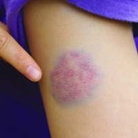
Wounds can be separated into open or closed wounds. In a closed wound the surface of the skin is intact, but the underlying tissues may be damaged. Examples of closed wounds are contusions, hematomas, or stage 1 pressure ulcers. With open wounds the skin is split or cracked, and the underlying tissues are exposed to the outside environment.


