Trigeminal Nerve
Original Editor - Saumya Srivastava
Top Contributors - Saumya Srivastava, Riccardo Ugrin, Wendy Walker, Kim Jackson, Vidya Acharya and Ahmed M Diab
Description[edit | edit source]
The Trigeminal Nerve (TNr) is the fifth cranial nerve. It is also represented as CN V. It is the largest of all the cranial nerves. It is also the most complex of all the cranial nerves as it has an extensive anatomic course. Knowing its course further helps in understanding the relationship between the brainstem, skull base, and facial area.
Trigeminal nerve is a mixed nerve - having both sensory and motor fibres. The origin of the trigeminal nerve is the annular protuberance at the limit of the cerebellar peduncles[1]. It originates from three sensory nuclei (mesencephalic, principal sensory, spinal nuclei of trigeminal nerve) and one motor nucleus (motor nucleus of the trigeminal nerve) extending from the midbrain to the medulla[2].
Distal Roots -[edit | edit source]
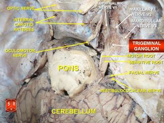
- The sensory nuclei merge to form the sensory root at the level of the pons and the motor nucleus continues to form the motor root.
- As a result the sensory root is large (made up of about 50 fascicles)[1], while the motor root is more slender (composed of six or seven fascicles)[1].These roots are analogous to the dorsal and ventral roots of the spinal cord[2]
- The sensory root then expands into the trigeminal ganglion, in the middle of the cranial fossa. The trigeminal ganglion is located lateral to the cavernous sinus, in a depression of the temporal bone, known as the trigeminal cave[2].
Peripheral Divisons of The TNr into Branches-[edit | edit source]
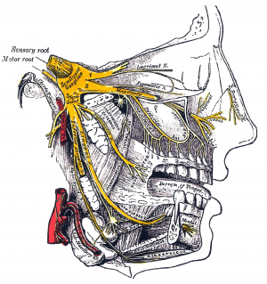
The TNr divides into 3 nerves distal to the trigeminal ganglion[3] -
- The Ophthalmic nerve (V1), passes forward in the lateral wall to the cavernous sinus. It then enters the orbit via the superior orbital fissure.
- The Maxillary nerve (V2), leaves the skull base through the foramen rotundum ossis sphenoidalis, inferolateral to the cavernous sinus. It then enters the pterygopalatine fossa, giving off several branches. The main trunk then continues anteriorly in the orbital floor and emerges onto the face as the infraorbital nerve
- The Mandibular nerve (V3), runs laterally along the skull base then exits the cranium by descending through the foramen ovale into the masticator space. The motor root of the trigeminal nerve bypasses the trigeminal ganglion and reunites with the mandibular nerve in the foramen ovale basis cranii. Hence the V3 contains both sensory and motor components.
Function[edit | edit source]
It is a mixed nerve - the sensory part of the nerve supplies the face (includes touch, pain, and temperature) and the motor part is for muscles of mastication[3]. The sensory information is sent forth through the main trigeminal nucleus and nuclei of the thalamus before it travels to the cerebral cortex and synapses in the post-central gyrus. Just like any other sensory information of the body, the information from the face crosses over (decussates) to the contralateral brain hemisphere[5].
Motor -[edit | edit source]
- The motor root of the trigeminal nerve bypasses the trigeminal ganglion and reunites with the mandibular nerve in the foramen ovale basis cranii.
- The motor root of the mandibular nerve innervates the four muscles of mastication:
- the mylohyoid,
- the anterior belly of digastric,
- the tensor muscle of the tympanic membranes, and
- tensor muscle of valum palatinum
Sensory[5] -[edit | edit source]
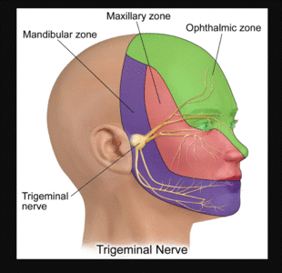
- The ophthalmic nerve is responsible for sensory innervation of the face and skull above the palpebral fissure as well as the eye and portions of the nasal cavity. It also contains sympathetic nerve fibers responsible for pupil dilation and supplies the ciliary body, iris, lacrimal gland, conjunctiva, and cornea. Additionally also supplies the superior portion of the nasal cavity, the frontal sinus, and even deeper structures including the dura mater and portions of the anterior cranial fossa.
- The maxillary nerve innervates portions of the nasal cavity, sinuses, maxillary teeth, palate, and the middle portion of the face and skull above the mouth and below the forehead.
- The sensory fibres of the mandibular nerve are responsible for pain and temperature information from the mandibular teeth, buccal mucosa, temporomandibular joint, the face below the territory of the maxillary nerve and the anterior two-thirds of the tongue; this is differentiated from taste which is produced by CN VII.
Clinical relevance[edit | edit source]
Assessment[edit | edit source]
Treatment[edit | edit source]
Resources[edit | edit source]
References[edit | edit source]
- ↑ 1.0 1.1 1.2 Barral JP, Croibier A. Trigeminal nerve. In:Barral JP, Croibier A editor(s). Manual Therapy for the Cranial Nerves. Churchill Livingstone, 2009, Pages 107-114,
- ↑ 2.0 2.1 2.2 Pazhaniappan N. The Trigeminal Nerve (CN V). Available from:https://teachmeanatomy.info/head/cranial-nerves/trigeminal-nerve/. (accessed 14 October 2020)
- ↑ 3.0 3.1 Kamal H.A.M, Toland J. Trigeminal Nerve Anatomy: Illustrated Using Examples of Abnormalities. American Journal of Roentgenology. 2001;176: 247-251.
- ↑ Trigeminal Nerve Anatomy - Cranial Nerve 5 Course and Distribution. Available from: http://www.youtube.com/watch?v=KZl3zdUAd-w
- ↑ 5.0 5.1 Huff T, Daly DT. Neuroanatomy, Cranial Nerve 5 (Trigeminal) [Updated 2020 Jul 31]. In: StatPearls [Internet]. Treasure Island (FL): StatPearls Publishing; 2020 Jan-.
Original Editor - Saumya Srivastava
Top Contributors - Saumya Srivastava, Riccardo Ugrin, Wendy Walker, Kim Jackson, Vidya Acharya and Ahmed M Diab


