Triceps brachii: Difference between revisions
(palpation) |
(Moved the Kenhub video to the resource section) |
||
| (16 intermediate revisions by 2 users not shown) | |||
| Line 4: | Line 4: | ||
'''Top Contributors''' - {{Special:Contributors/{{FULLPAGENAME}}}} | '''Top Contributors''' - {{Special:Contributors/{{FULLPAGENAME}}}} | ||
</div> | </div> | ||
== Description == | == Description == | ||
The | [[File:Triceps brachii muscle - animation02.gif|right|frameless]] | ||
The triceps brachii is a large, thick [[muscle]] on the dorsal part of the upper arm. It often appears as the shape of a horseshoe on the posterior aspect of the arm. The main function of the triceps is the extension of the [[Elbow|elbow joint]].<ref name=":0">Tiwana MS, Sinkler MA, Bordoni B. Anatomy, [https://www.statpearls.com/articlelibrary/viewarticle/30580/ Shoulder and Upper Limb, Triceps Muscle].Available:https://www.statpearls.com/articlelibrary/viewarticle/30580/ (accessed 30.12.2021)</ref> | |||
The Triceps brachii gets its name with tri referring to "three" muscle heads or points of origin (with Brachii referring to the arm). These include the: Medial head; Lateral head; Long head | |||
Image 1: Triceps brachii muscle: Long head red; Lateral head yellow; Medial head green | |||
=== | === Triceps brachii - Muscles === | ||
Long head | |||
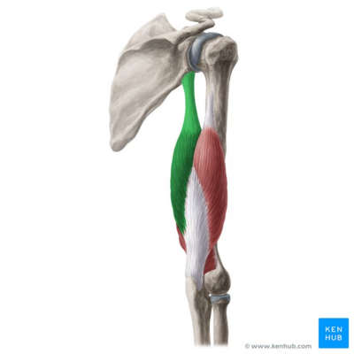
* [[File:Long head of triceps brachii muscle - Kenhub.png|alt=Long head of triceps brachii muscle (highlighted in green) - posterior view|right|frameless|Long head of triceps brachii muscle (highlighted in green) - posterior view|400x400px]]Origin: infraglenoid tubercle of the [[scapula]] | |||
* Insertion: Posterior surface of the olecranon process of the [[ulna]], capsule of the elbow joint and antebrachial [[fascia]]. | |||
* Action: Because it attaches to the scapula, the long head not only extends the elbow but will also have a small action on the [[Glenohumeral Joint|glenohumeral]] joint. With the arm adducted, the triceps muscle acts to hold the head of the humerus in the glenoid cavity. This action helps prevent any displacement of the humerus. The long head also assists with the extension and adduction of the arm at the shoulder joint. The lateral head is also active during extension of the forearm at the elbow joint when the forearm is supinated or pronated. | |||
* Innervation: [[radial nerve]] | |||
Image 2: Long head of triceps brachii muscle (highlighted in green) - posterior view <ref > Long head of triceps brachii (highlighted in green) - posterior view image - © Kenhub https://www.kenhub.com/en/library/anatomy/triceps-brachii-muscle </ref> | |||
[[File:Triceps .png|right|frameless]] | |||
Lateral Head | |||
* Origin: posterior aspect of the [[humerus]], superior to the radial groove | |||
* Insertion: Posterior surface of the olecranon process of the ulna, capsule of the elbow joint and antebrachial fascia. | |||
* Action: Strongest head of the three. It is active during extension of the forearm at the elbow joint when the forearm is supinated or pronated. | |||
* Innervation: radial nerve | |||
Medial head | |||
* Origin: posterior aspect of humerus, inferior to the radial groove | |||
* Insertion: Posterior surface of the olecranon process of the ulna, capsule of the elbow joint and antebrachial fascia. | |||
* Action: As the medial head does not attach to the scapula and therefore has no action on the glenohumeral joint, whether with stabilization or movement. However, the medial head is active during extension of the forearm at the elbow joint when the forearm is supinated or pronated. | |||
* | * Innervation: radial nerve<ref name=":0" /> | ||
* | Note: All the three heads of triceps brachii are innervated by the four branches of the [[radial nerve]] (C7, C8). However, according to the cadaveric study it is found that the medial head of triceps brachii is innervated by the [[Ulnar Nerve|ulnar nerve]].<ref>Bekler H, Wolfe VM, Rosenwasser MP. [https://www.ncbi.nlm.nih.gov/pmc/articles/PMC2600974/ A cadaveric study of ulnar nerve innervation of the medial head of triceps brachii.] Clinical orthopaedics and related research. 2009 Jan 1;467(1):235-8.</ref>. Some research reveals that the long head of triceps is actually innervated by the [[Axillary Nerve Injury|axillary nerve]].<ref>Triceps Brachii: Functional Anatomy Guide-The Definitive Guide to Triceps Brachii Anatomy, Exercises & Rehab[http://www.kingofthegym.com/triceps-brachii/ www.kingofthegym.com/triceps-brachii/](accessed 12 June 2018) | ||
</ref> | </ref> | ||
The muscle is supplied with [[oxygen]] and [[Nutrition|nutrients]] from the branches of the deep brachial [[Arteries|artery]].<ref>Triceps Anatomy, Origin & Function | Body Maps[https://www.healthline.com/human-body-maps/triceps#1 www.healthline.com/human-body-maps/triceps#1](accessed 13 June 2018)</ref> | |||
== | === Muscle Fibre Types === | ||
Muscle histology and average innervation ratios estimated from absolute mononeurons (MN) counts shows that the lateral head is used for movements requiring occasional high-intensity force, while the medial head enables more precise, low-force movements. | |||
# medial head was predominantly formed by small type I [[Muscle Fibre Types|muscle fibers]] and motor units (69 fibers/MN). | |||
# the lateral head comprised a great quantity of large type IIb fibers and motor units (179 fibers/MN) | |||
# the long head consisted of a more balanced mixture of fiber types and motor units (99 fibers/MN).<ref>Lucas‐Osma AM, Collazos‐Castro JE. [https://onlinelibrary.wiley.com/doi/abs/10.1002/cne.22123 Compartmentalization in the triceps brachii motoneuron nucleus and its relation to muscle architecture.] Journal of Comparative Neurology. 2009 Sep 20;516(3):226-39.</ref> | |||
== | == Physiotherapy == | ||
The triceps [[Reflexes|reflex]], elicited by sharply striking the triceps tendon, is often used to test the function of the [[Neurone|nerves]] of the arm. This reflex tests spinal nerves C6 and C7, predominately C7. The reflex is tested by abducting the patient's shoulder and elbow to 90 degrees. Then the triceps tendon is tapped using a reflex hammer just proximal to the olecranon. | |||
[[Axillary Nerve Injury|Axillary nerve damage]] can have an impact on the long head of the triceps brachii (LTB). Therefore, people with axillary nerve damage should undergo an assessment for the function of the LTB. If they demonstrate lost function, this shows a poor prognosis, and early repair at three months is recommended. | |||
The triceps muscle can undergo reinnervation through a distal nerve transfer. Commonly used nerves for reinnervation include the [[Flexor Carpi Ulnaris Muscle|flexor carpi ulnaris]] fascicle of the ulnar nerve and the posterior branch of the axillary nerve. Both of these nerves have been shown to recover the function of the triceps muscle. | |||
Ruptures of the triceps muscle are rare and typically only occur in [[Anabolic Steroid Abuse|anabolic steroid users]]. Distal [[Triceps tendon avulsion|triceps ruptures]] are also relatively uncommon. The injury typically occurs due to either a fall on an outstretched hand or a direct blow to the triceps tendon. The patient presents after a painful popping sensation with pain and swelling over the posterior elbow with the inability to extend the elbow against resistance. | |||
[[File:Tricep dip.jpeg|right|frameless|450x450px]] | |||
[[Strength Training|Strengthening exercises]]: The triceps brachii can undergo training in a variety of different ways. It can be worked out in isolation or with compound elbow extension movements. It can also be contracted statically to keep the arm straight against resistance. | |||
Examples | |||
# Isolation movements: lying triceps extensions, behind-the-back arm extensions, cable push-downs, and standing triceps "kickbacks." | |||
# The compound exercises: any pressing movements like [[Pushups|push-ups]], bench press, close grip bench press, tricep dips, and military presses. The closer the grip on these exercises, the more they isolate the triceps. The wider the grip, the more they work the outer chest. | |||
# Static contraction movements include pullovers, straight-arm pull-downs, and bent-over lateral raises, which are also used to build the deltoids and latissimus dorsi. | |||
Watch these short videos below for more ideas | |||
Strengthening{{#ev:youtube|t9WrFg4KfPo}}[[Stretching]]{{#ev:youtube|hSaqjF0dMMg}} | |||
=== Palpation === | |||
{{#ev:youtube|10cTxLvLbfE}} | |||
To palpate the three heads of the triceps have the patient in high sitting position and stand behind the patient | |||
# Palpation of medial head- Firstly, use a landmark to palpate the muscle. In this case, the landmark would be medial epicondyle of the humerus. The examiner will place his/her three fingers just above the medial epicondyle and instruct the patient to extend the elbow pushing downward force on the couch as if to lift himself or herself up. Finally palpate the medial head. | |||
# Palpation of long head- Palpate from medial condyle of the humerus up to the axilla posteriorly would be the area for long head of triceps. Just beneath the axilla posteriorly, the examiner will place his/her three fingers & instruct the patient to extend the elbow pushing downward. Finally, palpate the long head of the triceps. | |||
# Palpation of lateral head- In order to palpate for the lateral head, place three fingers to the postero-lateral side in the middle of the shaft of the humerus & instruct the patient to extend the elbow. | |||
# Anconeus- To palpate the anconeus, the landmark would be lateral condyle of the humerus to the proximal ulna & instruct the patient to extend the elbow. | |||
== Length test == | |||
{{#ev:youtube|aqylgBezTk8}} | |||
{{#ev:youtube| | == Myofascial Release Technique == | ||
{{#ev:youtube|DeHkYQ8mjEo}} | |||
== | == Resources == | ||
The 3 minute video is on the anatomy of Triceps <ref > Triceps brachii muscle video - © Kenhub https://www.kenhub.com/en/library/anatomy/triceps-brachii-muscle</ref> | |||
{{#ev:youtube|R-TxrDzYSdM}} | |||
{{#ev:youtube| | |||
== References== | == References== | ||
Latest revision as of 18:16, 17 March 2023
forOriginal Editor - Esraa Mohamed Abdullzaher
Top Contributors - Mandeepa Kumawat, Esraa Mohamed Abdullzaher, Lucinda hampton, Joao Costa, Sai Kripa, Kim Jackson, Nikhil Benhur Abburi, Kirenga Bamurange Liliane and WikiSysop
Description[edit | edit source]

The triceps brachii is a large, thick muscle on the dorsal part of the upper arm. It often appears as the shape of a horseshoe on the posterior aspect of the arm. The main function of the triceps is the extension of the elbow joint.[1]
The Triceps brachii gets its name with tri referring to "three" muscle heads or points of origin (with Brachii referring to the arm). These include the: Medial head; Lateral head; Long head
Image 1: Triceps brachii muscle: Long head red; Lateral head yellow; Medial head green
Triceps brachii - Muscles[edit | edit source]
Long head
- Origin: infraglenoid tubercle of the scapula
- Insertion: Posterior surface of the olecranon process of the ulna, capsule of the elbow joint and antebrachial fascia.
- Action: Because it attaches to the scapula, the long head not only extends the elbow but will also have a small action on the glenohumeral joint. With the arm adducted, the triceps muscle acts to hold the head of the humerus in the glenoid cavity. This action helps prevent any displacement of the humerus. The long head also assists with the extension and adduction of the arm at the shoulder joint. The lateral head is also active during extension of the forearm at the elbow joint when the forearm is supinated or pronated.
- Innervation: radial nerve

Image 2: Long head of triceps brachii muscle (highlighted in green) - posterior view [2]

Lateral Head
- Origin: posterior aspect of the humerus, superior to the radial groove
- Insertion: Posterior surface of the olecranon process of the ulna, capsule of the elbow joint and antebrachial fascia.
- Action: Strongest head of the three. It is active during extension of the forearm at the elbow joint when the forearm is supinated or pronated.
- Innervation: radial nerve
Medial head
- Origin: posterior aspect of humerus, inferior to the radial groove
- Insertion: Posterior surface of the olecranon process of the ulna, capsule of the elbow joint and antebrachial fascia.
- Action: As the medial head does not attach to the scapula and therefore has no action on the glenohumeral joint, whether with stabilization or movement. However, the medial head is active during extension of the forearm at the elbow joint when the forearm is supinated or pronated.
- Innervation: radial nerve[1]
Note: All the three heads of triceps brachii are innervated by the four branches of the radial nerve (C7, C8). However, according to the cadaveric study it is found that the medial head of triceps brachii is innervated by the ulnar nerve.[3]. Some research reveals that the long head of triceps is actually innervated by the axillary nerve.[4]
The muscle is supplied with oxygen and nutrients from the branches of the deep brachial artery.[5]
Muscle Fibre Types[edit | edit source]
Muscle histology and average innervation ratios estimated from absolute mononeurons (MN) counts shows that the lateral head is used for movements requiring occasional high-intensity force, while the medial head enables more precise, low-force movements.
- medial head was predominantly formed by small type I muscle fibers and motor units (69 fibers/MN).
- the lateral head comprised a great quantity of large type IIb fibers and motor units (179 fibers/MN)
- the long head consisted of a more balanced mixture of fiber types and motor units (99 fibers/MN).[6]
Physiotherapy[edit | edit source]
The triceps reflex, elicited by sharply striking the triceps tendon, is often used to test the function of the nerves of the arm. This reflex tests spinal nerves C6 and C7, predominately C7. The reflex is tested by abducting the patient's shoulder and elbow to 90 degrees. Then the triceps tendon is tapped using a reflex hammer just proximal to the olecranon.
Axillary nerve damage can have an impact on the long head of the triceps brachii (LTB). Therefore, people with axillary nerve damage should undergo an assessment for the function of the LTB. If they demonstrate lost function, this shows a poor prognosis, and early repair at three months is recommended.
The triceps muscle can undergo reinnervation through a distal nerve transfer. Commonly used nerves for reinnervation include the flexor carpi ulnaris fascicle of the ulnar nerve and the posterior branch of the axillary nerve. Both of these nerves have been shown to recover the function of the triceps muscle.
Ruptures of the triceps muscle are rare and typically only occur in anabolic steroid users. Distal triceps ruptures are also relatively uncommon. The injury typically occurs due to either a fall on an outstretched hand or a direct blow to the triceps tendon. The patient presents after a painful popping sensation with pain and swelling over the posterior elbow with the inability to extend the elbow against resistance.

Strengthening exercises: The triceps brachii can undergo training in a variety of different ways. It can be worked out in isolation or with compound elbow extension movements. It can also be contracted statically to keep the arm straight against resistance.
Examples
- Isolation movements: lying triceps extensions, behind-the-back arm extensions, cable push-downs, and standing triceps "kickbacks."
- The compound exercises: any pressing movements like push-ups, bench press, close grip bench press, tricep dips, and military presses. The closer the grip on these exercises, the more they isolate the triceps. The wider the grip, the more they work the outer chest.
- Static contraction movements include pullovers, straight-arm pull-downs, and bent-over lateral raises, which are also used to build the deltoids and latissimus dorsi.
Watch these short videos below for more ideas
Strengthening
Palpation[edit | edit source]
To palpate the three heads of the triceps have the patient in high sitting position and stand behind the patient
- Palpation of medial head- Firstly, use a landmark to palpate the muscle. In this case, the landmark would be medial epicondyle of the humerus. The examiner will place his/her three fingers just above the medial epicondyle and instruct the patient to extend the elbow pushing downward force on the couch as if to lift himself or herself up. Finally palpate the medial head.
- Palpation of long head- Palpate from medial condyle of the humerus up to the axilla posteriorly would be the area for long head of triceps. Just beneath the axilla posteriorly, the examiner will place his/her three fingers & instruct the patient to extend the elbow pushing downward. Finally, palpate the long head of the triceps.
- Palpation of lateral head- In order to palpate for the lateral head, place three fingers to the postero-lateral side in the middle of the shaft of the humerus & instruct the patient to extend the elbow.
- Anconeus- To palpate the anconeus, the landmark would be lateral condyle of the humerus to the proximal ulna & instruct the patient to extend the elbow.
Length test[edit | edit source]
Myofascial Release Technique[edit | edit source]
Resources[edit | edit source]
The 3 minute video is on the anatomy of Triceps [7]
References[edit | edit source]
- ↑ 1.0 1.1 Tiwana MS, Sinkler MA, Bordoni B. Anatomy, Shoulder and Upper Limb, Triceps Muscle.Available:https://www.statpearls.com/articlelibrary/viewarticle/30580/ (accessed 30.12.2021)
- ↑ Long head of triceps brachii (highlighted in green) - posterior view image - © Kenhub https://www.kenhub.com/en/library/anatomy/triceps-brachii-muscle
- ↑ Bekler H, Wolfe VM, Rosenwasser MP. A cadaveric study of ulnar nerve innervation of the medial head of triceps brachii. Clinical orthopaedics and related research. 2009 Jan 1;467(1):235-8.
- ↑ Triceps Brachii: Functional Anatomy Guide-The Definitive Guide to Triceps Brachii Anatomy, Exercises & Rehabwww.kingofthegym.com/triceps-brachii/(accessed 12 June 2018)
- ↑ Triceps Anatomy, Origin & Function | Body Mapswww.healthline.com/human-body-maps/triceps#1(accessed 13 June 2018)
- ↑ Lucas‐Osma AM, Collazos‐Castro JE. Compartmentalization in the triceps brachii motoneuron nucleus and its relation to muscle architecture. Journal of Comparative Neurology. 2009 Sep 20;516(3):226-39.
- ↑ Triceps brachii muscle video - © Kenhub https://www.kenhub.com/en/library/anatomy/triceps-brachii-muscle

